

Written by: Dr. Akash Chandawarkar, Board Certified Plastic Surgeon, Mirror Plastic Surgery | Last updated: September 9, 2026
Key Takeaways
- Dermal fillers can cause scarring or long-term tissue damage, but peer-reviewed evidence shows these outcomes are rare, occurring in well under 1% of procedures with trained injectors.
- Vascular occlusion leading to tissue necrosis occurs in approximately 0.1%–0.3% of filler procedures, with higher risk in specific anatomical zones such as the glabella, nasal dorsum, and forehead.
- Histologically confirmed granulomas occur in roughly 0.02%–0.4% of cases, while permanent fillers like PMMA carry disproportionately higher long-term risks compared to modern biodegradable options.
- Injector anatomical expertise is the most important modifiable risk factor across all complication categories, so provider selection is the primary risk-reduction strategy.
- Provider selection is the key step patients control to lower the chance of serious complications.
Direct Answer: Long-Term Scarring and Tissue Damage Risk
Dermal fillers can cause scarring or tissue damage in rare cases, yet with an experienced, board-certified injector, the risk stays very low.1 Vascular events occur in approximately 0.1%–0.3% of procedures, and granulomas appear in a small fraction of patients.1 Most complications remain minor and treatable. Choosing a provider with deep anatomical knowledge and a safety-first approach has the greatest impact on your outcome.
The distinction between possible and probable matters for real-world decision-making. A 2026 systematic review in Aesthetic Plastic Surgery found low complication rates for modern fillers, largely because they are reversible. Permanent fillers such as PMMA and silicone are disproportionately associated with chronic inflammatory, vascular, and deforming complications. The same review reported intra-arterial injection with subsequent necrosis in 0.1%–0.3% of filler procedures, more commonly in the lip, forehead, and nasolabial fold. Modern fillers, which Mirror Plastic Surgery uses almost exclusively, show documented long-term complication rates below 0.5% across categories.1 Risk rises sharply with permanent fillers, injector inexperience, or off-label products.
Mechanisms of Scarring and Tissue Damage: Insights From 2026 Studies
Four documented pathways explain how filler complications can lead to scarring or lasting tissue change.
Vascular Occlusion: The Emergency Pathway
When filler enters or compresses a blood vessel, blood flow drops or stops, and tissue loses oxygen. A 2026 prospective case series by Olivares et al. in the Journal of Cosmetic Dermatology described this as a cascade of endothelial dysfunction, microvascular collapse, inflammatory amplification, and cellular hypoxia that can progress to irreversible necrosis. Inadequately treated ischemic events often result in contractile scarring through TGF-β–mediated signaling pathways that promote pathological fibrosis. Incidence is estimated at 1 in 2,000 to 1 in 10,000 procedures, with a 30-fold increase in documented cases between 2000 and 2020.1
Granuloma Formation: The Immune Pathway
The body can mount a foreign-body reaction to filler material and form small lumps of scar-like granulation tissue. A 2026 case series by Spivak found that delayed complications appeared a mean of 10.8 months after injection, with fibrotic induration present in 73.3% of cases and inflammatory nodules in 66.7%. Histopathology revealed foreign-body granulomas containing multinucleated giant cells. A 2026 narrative review in the Journal of Cosmetic Dermatology estimates that histologically confirmed granulomas remain uncommon overall.
Repeated Needle Trauma: The Cumulative Pathway
Each injection creates a micro-track through tissue. A 2026 scoping review in the Brazilian Dental Journal found that bruising (56.4%), ischemia (45.4%), and edema (41.8%) were the most frequently reported adverse reactions across 55 studies. Individual tracks usually stay insignificant. Repeated superficial placement over many years may theoretically contribute to fibrotic change, although no 2026 study has shown clinically significant scarring from this mechanism alone.
Delayed Inflammatory Reactions: The Late-Onset Pathway
The 2026 Spivak case series documented that infections (26.7%) and vaccinations (13.3%) triggered delayed complications in susceptible patients.1 These findings suggest that immune stimuli can provoke late inflammatory responses to HA fillers. Ultrasound showed hypoechoic deposits with fibrotic halos, confirming that HA fillers can trigger lasting tissue responses rather than behaving as fully inert materials.
Long-Term Tissue Fate: What Happens To Fillers Over Time
A 2026 MRI study by Issa et al. in Plastic and Reconstructive Surgery Global Open challenged long-held assumptions about filler longevity. Using 3D MRI on 14 patients, researchers found that detectable filler volume averaged 2.8 times the amount initially injected, with filler present up to 180 months (15 years) post-injection. The tear trough region showed the greatest proportional increase in filler thickness.
A 2026 narrative review by Alizadeh et al. notes that MRI visualizes a combination of injected filler, bound water, and newly formed collagen. Apparent persistence does not mean the original product remains intact. Gels have documented biostimulatory properties that promote collagen deposition, with up to a 12-fold increase in type I procollagen gene expression described.
Clinical implications for patients include:
- Fillers are reversible in many cases. Hyaluronidase dissolves HA fillers, with reversal documented in 11 published case series including 341 patients.
- Biostimulatory fillers are not enzyme-reversible. Calcium hydroxylapatite complications rely more heavily on prevention and supportive care, because no enzymatic reversal exists.
- Filler persistence does not automatically signal higher complication risk. A 2026 prospective cohort study in JPRAS Open followed 60 patients receiving a low-modification filler over 24 months and found zero granulomas, zero delayed nodules, and no persistent inflammatory reactions.
Risk Factor Analysis: What the Data Shows About Predictors
A 2026 systematic review in Aesthetic Plastic Surgery concluded that anatomic injection site, technique, and filler permanence are the principal determinants of irreversible injury. Injected volume alone plays a smaller role. The evidence highlights several specific risk factors.
The first and most dangerous risk factor is the anatomical zone being treated. High-risk anatomical zones. The glabella, nasal dorsum, and forehead carry the highest vascular occlusion risk due to direct anastomoses with the ophthalmic artery. Injections in these regions can lead to skin necrosis or permanent vision loss in rare cases. The Olivares et al. study identified the glabella, nasal dorsum, nasal tip, alar region, nasolabial fold, lips, midface, and temporal fossa as high-risk vascular territories.
Equally important is the choice of product. Filler type selection. Permanent fillers (PMMA, silicone) are disproportionately associated with chronic complications. The 2026 Aesthetic Plastic Surgery review reports granuloma rates of 1–2% for PMMA fillers and approximately 6% for collagen injections, far higher than the low rates seen with HA fillers. The FDA has not approved injectable silicone or any injectable fillers for body contouring.
Technique and training then shape how those products behave in the face. Injector technique and training. The Olivares et al. study found significantly lower incidence of ischemic events when cannulas were used compared to needles. A 2026 retrospective study found that over half of filler complications presenting to a university dermatology department involved either uncertified products or injections by non-medical practitioners. The 2026 Aesthetic Plastic Surgery review identified practitioner lack of expertise, lack of informed consent, and delayed diagnosis as common factors in malpractice cases. Median awards in these cases ranged from $262,000 to $600,000.
Finally, individual health and history influence risk. Patient-specific factors. A 2026 expert consensus in the Journal of Drugs in Dermatology identified patient history, product characteristics, procedural technique, and post-procedure care as the four risk factor categories that require attention. Effective risk reduction addresses all four categories together.
Expert Analysis: Why Injector Expertise Matters Most
The research converges on a clear conclusion: the injector’s anatomical knowledge and technical skill are the most powerful modifiable risk factors. A 2026 scoping review in the Brazilian Dental Journal emphasized that injectors must recognize complication signs early and manage them confidently. The 2026 Journal of Cosmetic Dermatology review concluded that with appropriate training and safety algorithms, most filler complications can be treated effectively.
This evidence directly shapes the safety philosophy at Mirror Plastic Surgery. Dr. Akash Chandawarkar is a board-certified plastic surgeon with fellowship training in aesthetic surgery at the Manhattan Eye Ear & Throat Hospital. Because he personally performs all injections, every treatment benefits from a surgeon’s command of facial and subdermal anatomy, the expertise the literature identifies as the primary protective factor against serious complications.
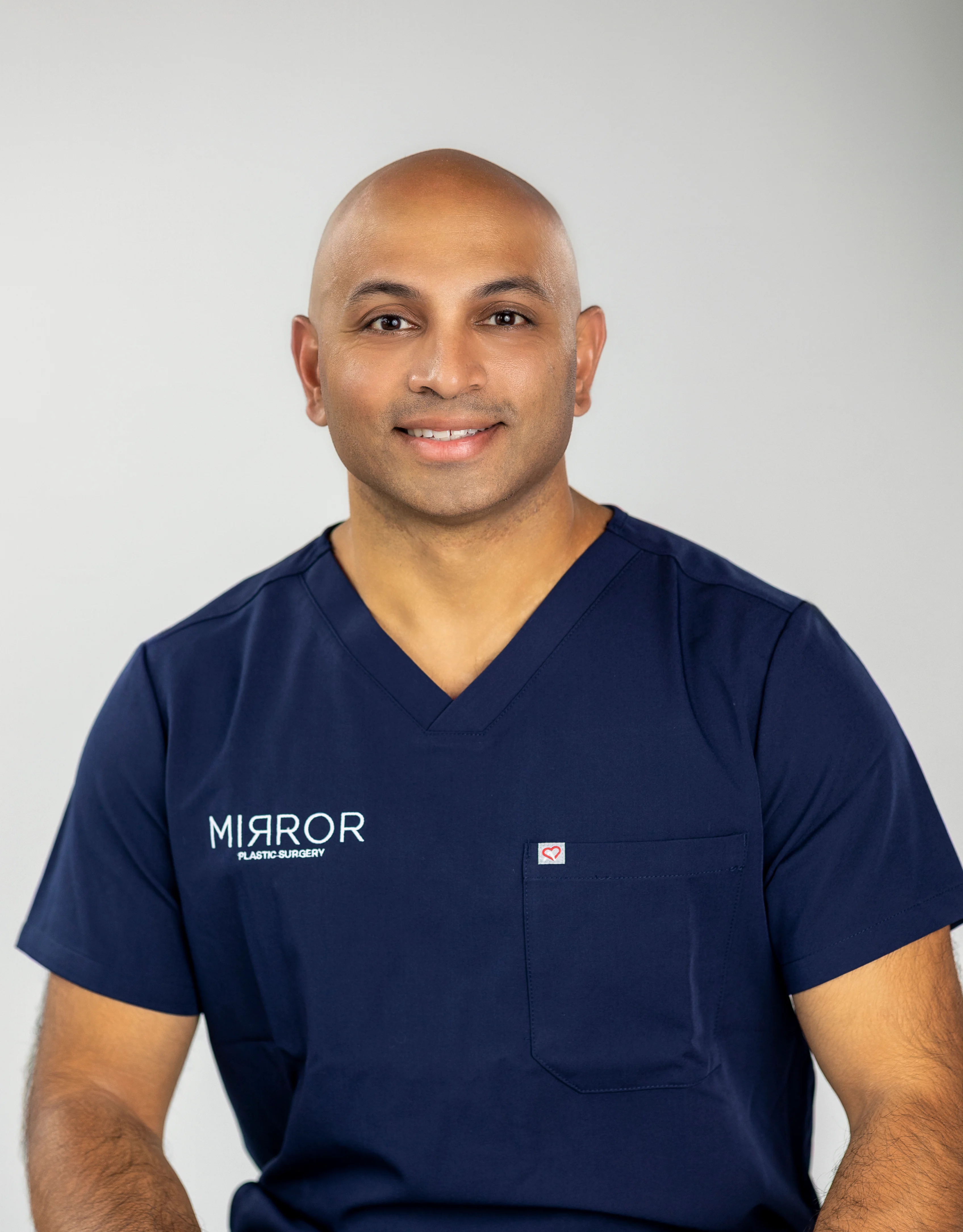
For patients, this translates into several concrete safety advantages:
- Board certification matters. Plastic surgeons complete years of anatomy training that non-physician injectors do not receive.
- Complication management capability matters. A surgeon who can recognize and treat vascular occlusion immediately, with hyaluronidase, nitroglycerin paste, and hyperbaric oxygen referral pathways, can prevent permanent damage.
- Conservative treatment philosophy matters. The 2026 Issa et al. MRI findings support conservative dosing and long-term procedural planning, because HA fillers can expand and persist longer than expected.
Discuss your filler goals with Dr. Chandawarkar to receive a safety-first treatment plan grounded in 2026 evidence.
Planning Future Procedures With Existing Fillers
The 2026 Aesthetic Plastic Surgery review notes that filler migration occurs in up to 1% of cases and tissue atrophy in fewer than 1% of patients, particularly with high-volume fillers or repetitive treatments. A 2026 review by Alizadeh et al. found that in a series of lip augmentation patients, only 2 of 27 treated solely with HA gels had documented migration.
These findings guide planning for future procedures:
- HA fillers can be dissolved before surgery. Many surgeons recommend dissolving filler several weeks before a facelift to restore more natural anatomy.
- Biostimulatory fillers require advance planning. Products such as Radiesse are not enzyme-reversible, so surgical planning must account for their presence.
- Conservative dosing preserves future options. The MRI evidence that apparent filler volume can exceed injected volume supports a “less is more” philosophy from the start.
Evidence-Based Recommendations For Patients
Based on the 2026 research literature and expert consensus guidelines, patients considering dermal fillers can take several practical steps.
Verify injector credentials. Seek a board-certified plastic surgeon or dermatologist with documented injection experience. The 2026 Aesthetic Plastic Surgery review identifies insufficient understanding of facial anatomy, wrong filler selection, poor technique, and poor diagnosis or management as causes of permanent complications.
Ask about complication protocols. Confirm that your provider has hyaluronidase on hand and a clear plan for recognizing and responding to vascular events. The 2026 Journal of Cosmetic Dermatology review emphasizes that immediate high-dose hyaluronidase is the standard of care for vascular occlusion.
Request a comprehensive consultation. A thorough assessment that evaluates your full face, medical history, and aesthetic goals, not just the injection site, helps identify risk factors before treatment begins. At Mirror Plastic Surgery, Dr. Chandawarkar’s concierge-level consultations routinely extend up to an hour for this reason.
Choose reversible fillers for safety. The 2026 Aesthetic Plastic Surgery review states that reversible fillers are preferred for most facial areas because they can be dissolved with hyaluronidase. Permanent fillers should be used with extreme caution or avoided.
Frequently Asked Questions
Can You Get Scar Tissue From Fillers?
Scar-like tissue can form after fillers, but it remains uncommon. Granulomas, which represent a type of scar-like tissue formed by the immune response to filler, occur at low rates in published series. Fibrotic induration was the most common finding (73.3%) in a 2026 case series of delayed filler complications. These reactions are usually treatable with hyaluronidase and intralesional corticosteroids. Permanent fillers such as PMMA carry substantially higher granuloma rates, which is one reason Mirror Plastic Surgery prioritizes reversible formulations for facial treatments.
Does Filler Ever Fully Leave Your Body?
Fillers are biodegradable and eventually absorb, yet 2026 MRI research detected filler-related tissue changes up to 15 years post-injection. The same study found that MRI-measured volume averaged 2.8 times the injected amount, suggesting that fillers stimulate collagen production that persists beyond the product itself. This pattern reflects the biostimulatory properties of fillers and supports conservative dosing and long-term planning. Biostimulatory fillers like Radiesse are designed to stimulate lasting collagen and are not enzyme-reversible, so they require a different planning approach.
What Is the Riskiest Place for Fillers?
The glabella, nasal dorsum, and forehead carry the highest vascular occlusion risk because of direct arterial connections to the ophthalmic artery. Injections in these regions can lead to skin necrosis or vision loss in rare cases. The nasolabial fold and forehead are also associated with cerebral embolic events. The lips, midface, and temporal fossa are additional high-risk vascular territories. In all of these zones, injector anatomical expertise and the use of cannulas over needles provide key protection.
Do Fillers Affect Future Facelifts or Other Procedures?
Fillers can create scar tissue that may complicate surgical dissection, and experienced surgeons account for this during planning. HA fillers can be dissolved with hyaluronidase before surgery, while biostimulatory fillers cannot. Migration remains rare with HA gels when properly placed, and one review found migration in only 2 of 27 patients treated solely with HA gels. Conservative dosing and precise placement help preserve future surgical options, which is why Dr. Chandawarkar emphasizes a “less is more” philosophy from the first treatment.
How Do I Know If My Injector Is Qualified Enough?
Board certification in plastic surgery or dermatology provides a strong baseline credential. Beyond that, ask whether the provider personally performs every injection, whether they have hyaluronidase on hand for emergencies, and whether they conduct a comprehensive facial assessment before recommending any treatment. A provider who spends time understanding your anatomy, medical history, and long-term goals, rather than moving quickly to product recommendations, reflects the safety-first philosophy that the 2026 literature consistently identifies as most protective.
Conclusion: Making an Evidence-Based Filler Choice
The 2026 research literature shows that dermal fillers can cause scarring or tissue damage in rare cases, yet the overall risk remains low. Severe complications from hyaluronic acid fillers occur at rates below 0.5% (for example, 0.0041% for serious complications in a large study), while minor injection-site reactions and clinically meaningful nodules or biofilm can occur at higher rates, up to 5% depending on product and technique. Injector expertise, anatomical knowledge, careful technique, and appropriate product selection consistently emerge as the decisive protective factors.
At Mirror Plastic Surgery in St. Petersburg, FL, Dr. Akash Chandawarkar embodies the safety-first approach described in the research. As a board-certified plastic surgeon with fellowship training in aesthetic surgery, he personally performs every injection with a surgeon’s command of facial anatomy. His concierge-level consultations include a comprehensive top-to-bottom assessment that identifies risk factors before treatment begins, a standard that high-volume clinics rarely match.
If you are considering dermal fillers, the provider you choose is the most important decision you will make. Start your consultation with Ellie today to discuss your goals and receive a personalized, evidence-based treatment plan from a surgeon whose qualifications match the risk profile the literature describes.
Contact: Phone 727-361-6515 | Email hello@mirrorplasticsurgery.com | 780 4th Ave S, St. Petersburg, FL 33701
Disclaimer: Results may vary from person to person. Editorial content, before and after images, and patient testimonials do not constitute a guarantee of specific results.
1 Results may vary from person to person. Editorial content, before and after images, and patient testimonials do not constitute a guarantee of specific results.