

Written by: Dr. Akash Chandawarkar, Board Certified Plastic Surgeon, Mirror Plastic Surgery | Last updated: September 9, 2026
What This Dermal Filler Risk Report Covers
- This report reviews dermal filler risks, from common side effects to rare complications, and explains how expert injectors reduce those risks.
Understanding The Data On Filler Complications
Published complication rates underestimate real-world risk. The FDA notes that its passive MAUDE surveillance system captures only an estimated 1–5% of actual complications, so reported numbers represent only a fraction of events.
Peer-reviewed data provides a useful baseline:
- Over 90% of adverse events from hyaluronic acid fillers are mild and transient, such as injection-site redness, swelling, and bruising.1
- A 2026 scoping review in the Brazilian Dental Journal covering 55 studies found bruising reported in 56.4% of studies, ischemia in 45.4%, and edema in 41.8%.1
- Serious complications remain uncommon. Observational studies report vascular occlusion in approximately 1 in 2,000 to 1 in 10,000 procedures.1
These figures frame the discussion that follows and highlight both the frequency of events and the limits of current reporting systems.
Discuss your risk profile with a board-certified plastic surgeon at Mirror Plastic Surgery before committing to any filler treatment.
Common Side Effects After Filler Injections
Most patients experience predictable, short-term reactions after filler. These effects are normal physiological responses and should be reviewed in every consultation.
- Bruising is particularly frequent in highly vascular areas like the lips and under-eyes and typically resolves within 1–2 weeks.1
- Swelling peaks within 1–2 days and subsides over several days. It is more pronounced in the lips because of dense blood supply and loose connective tissue.1
- Redness, tenderness, and itching at injection sites usually fade within a few days.
A 2026 narrative review in the Journal of Cosmetic Dermatology confirms these as the most common minor adverse events. While temporary and manageable, these side effects highlight the need to schedule treatments with enough recovery time, especially before important events.
Rare But Serious Complications: Vascular Occlusion, Necrosis, And Blindness
Rare complications carry serious consequences and require careful attention from both patients and injectors.
- Vascular occlusion occurs when filler enters or compresses a blood vessel and blocks blood flow. The incidence matches the rate mentioned earlier, with a 30-fold increase in reported cases between 2000 and 2020.
- Skin necrosis refers to tissue death from prolonged ischemia and was reported in 29.1% of studies in the 2026 Brazilian Dental Journal scoping review.1
- Blindness remains extremely rare but devastating. Approximately 190 cases were reported worldwide between 1906 and 2019, mostly from autologous fat injections.1
The highest-risk zones include the glabella, nose, nasolabial folds, and forehead, which connect directly to the ophthalmic artery. An injector with a surgeon’s level of vascular knowledge and clear emergency protocols, including on-site hyaluronidase, can prevent many of these events or limit damage.
As Dr. Chandawarkar explains, the outcome often depends on how quickly the injector recognizes and treats a vascular event in the first minutes.
Long-Term Side Effects Of Repeated Dermal Filler Use
Long-term effects shape how the face ages with filler and often influence decisions about future treatment or dissolution.
- Filler migration occurs when product moves from the intended site into nearby tissue, causing visible distortion. MRI studies show hyaluronic acid filler persisting in facial tissues for 2 to 15 years after injection, and migration can appear months or years later.1
- Granulomas and delayed nodules are inflammatory lumps that may appear weeks to years after treatment, with an estimated incidence of 0.02%–0.4% for modern hyaluronic acid fillers.1
- The “overfilled” or “pillow face” look develops gradually after years of small additions, often without anyone noticing the exact point when balance is lost.
- Persistent edema involves malar or periorbital swelling that can last for months, particularly after tear-trough injections, and aligns with the 11% rate cited in the risk table below.1
- Residual filler complicating future surgery can alter tissue planes and obscure landmarks during facelift or blepharoplasty, making surgery more complex.
Hyaluronic acid fillers do not always fully dissolve on the manufacturer’s advertised timeline. Multiple MRI studies confirm persistence at 2–15 years. This persistence matters for patients who receive repeated treatments over many years and helps explain the growing interest in filler dissolution.
Why More Patients Are Choosing To Dissolve Their Fillers
Elective filler dissolution has become a visible trend in aesthetics. Market data from major practices shows a rise in elective hyaluronidase procedures, where patients seek removal of accumulated product rather than treatment of acute complications.
Common reasons for dissolution include:
- Dissatisfaction with results, such as lips that feel heavy, a midface that looks overfilled, or under-eyes that appear puffy.
- Filler migration, including visible product above the lip border (“filler mustache”) or in unintended areas.
- Shifting aesthetic preferences, as many people move from maximal volume toward subtle, natural results.
- “Filler fatigue”, the feeling of dependence on filler after years of maintenance treatments.
Hyaluronic acid fillers can be dissolved with hyaluronidase, yet the process has real tradeoffs. As Dr. Paul Nassif explained in the Los Angeles Times, the enzyme affects both injected filler and the body’s natural hyaluronic acid, so aggressive or repeated use can thin natural tissue. Dissolution often requires multiple sessions, and non-hyaluronic acid fillers like Radiesse or Sculptra cannot be dissolved.
Explore your options for filler dissolution or a conservative new plan with a board-certified plastic surgeon who understands both the risks and the aesthetics.
The Riskiest Places To Inject Dermal Fillers
Injection sites near the ophthalmic artery carry the highest stakes. The table below summarizes risk by area, with supporting citations.
| Treatment Area | Primary Risks | Risk Level |
|---|---|---|
| Nose (Non-Surgical Rhinoplasty) | Vascular compromise, skin necrosis, blindness, with dorsal nasal injection significantly associated with blindness in FDA MAUDE analysis | Highest |
| Under-Eyes (Tear Trough) | Bruising, Tyndall effect, malar edema in up to 11% of cases, vascular occlusion, and off-label use of all hyaluronic acid fillers in this area | High |
| Glabella | Vascular occlusion with retrograde embolization to the ophthalmic artery and blindness, with markedly higher risk in this zone | High |
| Lips | Swelling, lumps, migration above the vermilion border, and vascular risk from labial arteries, with bruising particularly frequent due to dense vascular supply | Moderate–High |
| Cheeks | Nerve injury, migration, persistent edema, with cheek identified as the most common adverse-event site (43%) in FDA MAUDE analysis | Moderate |
The 2025 ASPS and AAD position statements recommend that periocular injections be performed by a board-certified plastic surgeon, dermatologist, or oculoplastic surgeon with advanced anatomical training.
How A Board-Certified Plastic Surgeon Reduces Filler Risks
Injector training and experience strongly influence safety. A 2026 retrospective study of filler complications found that over half involved uncertified products or injections by non-medical practitioners. The Harley Street Institute white paper estimates that practitioners need about 3.85 years and 786 filler injections to reach “advanced” competency.
Dr. Akash Chandawarkar practices at this advanced level. He is Harvard-educated, Johns Hopkins-trained, board-certified by the American Board of Plastic Surgery, and fellowship-trained in aesthetic surgery at MEETH/Lenox Hill Hospital. His surgical command of facial and subdermal anatomy, the same expertise required for facelift surgery, guides every injection.
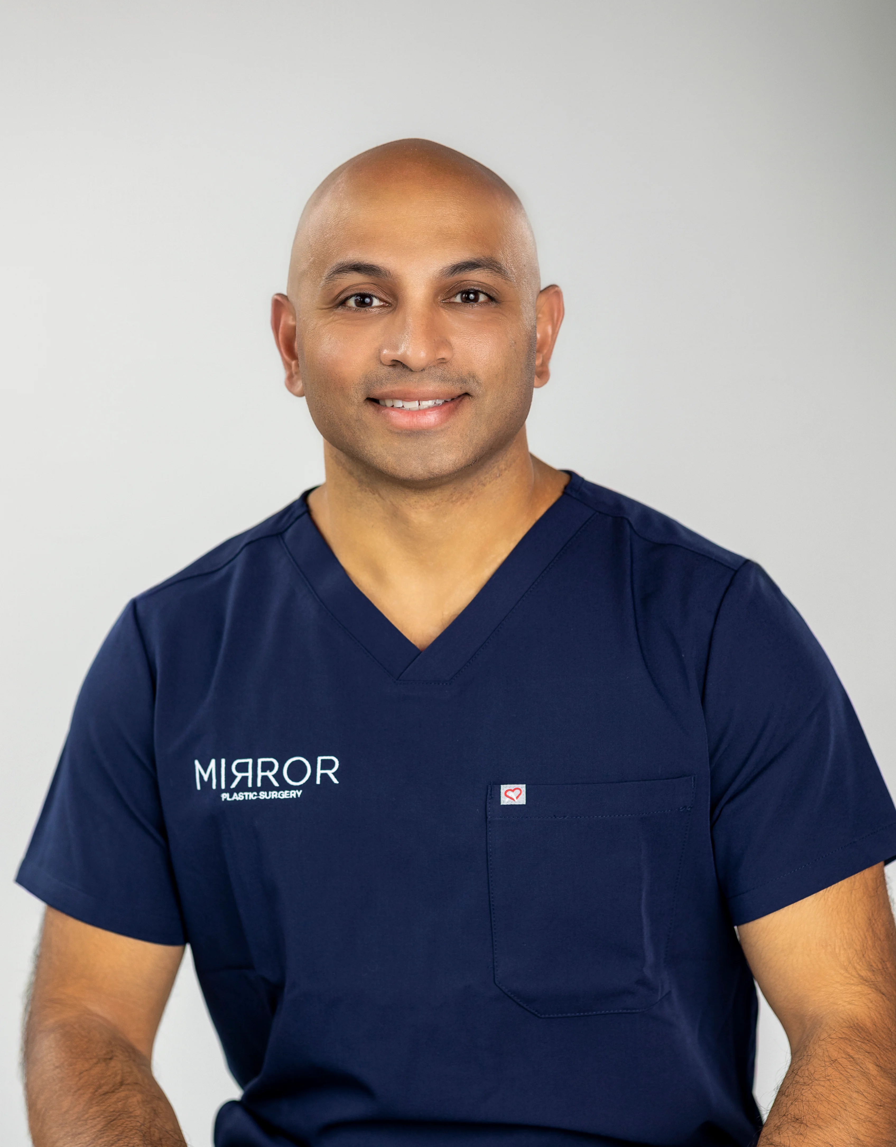
This surgical background shapes his philosophy of “safety first, function second, aesthetics third.” In practice, that means hour-long consultations, a top-to-bottom facial assessment, and honest conversations about whether fillers are appropriate for each patient.
How To Choose A Qualified Filler Provider
Patients can reduce risk by using a simple checklist when evaluating injectors.
- Verify board certification. Confirm certification by the American Board of Plastic Surgery or American Board of Dermatology through ABMS.org.
- Ask about emergency protocols. Ask whether the practice keeps hyaluronidase on-site and how they handle vascular complications. The FDA recommends that any practice injecting hyaluronic acid fillers maintain stocked, in-date hyaluronidase for emergency reversal.
- Request before-and-after photos of real patients rather than generic marketing images.
- Ask about their approach to natural results. A provider who recommends conservative volume and a full-face assessment before treating a single area is prioritizing safety.
- Avoid “filler mills”, where non-physicians perform high-volume injections in rushed, one-size-fits-all appointments.
Conclusion: Making A Thoughtful Decision About Dermal Fillers
Dermal fillers offer real benefits but carry meaningful downsides. Most complications are mild and short-lived, and many serious events are preventable when an expert injector follows strict safety protocols. The rise of filler dissolution reflects a broader shift toward restraint and natural results, which aligns with Dr. Chandawarkar’s safety-first, anatomy-driven approach.
For women in the St. Petersburg and Tampa area considering fillers, the evidence highlights one theme: the injector’s anatomical expertise shapes both safety and aesthetics. Schedule your consultation today to learn whether dermal fillers fit your goals and how to pursue subtle, harmonious results as safely as possible.
Frequently Asked Questions
What Are The Most Common Side Effects Of Dermal Fillers?
The most common side effects are mild and temporary. Bruising occurs frequently in highly vascular areas such as the lips and under-eyes and usually resolves within one to two weeks. Swelling peaks within one to two days and settles over several days, especially in the lips because of their blood supply and tissue structure. Redness, tenderness, and itching at injection sites typically fade within a few days. These reactions are expected physiological responses and should be reviewed during your consultation. Scheduling treatment at least two to three weeks before major events gives these effects time to resolve.
Does Filler Ever Fully Leave Your Body?
Hyaluronic acid filler can persist much longer than product brochures suggest. MRI and ultrasound studies published between 2022 and 2025 have repeatedly shown hyaluronic acid filler remaining in facial tissues for the 2–15 year window documented earlier. A 2024 study in Plastic and Reconstructive Surgery reviewed MRI scans of 33 patients who received midface hyaluronic acid filler and found residual product in every patient, including those who had not been injected for five to fifteen years. The visible effect softens as the filler degrades and binds less water, yet the material itself can remain. Non-hyaluronic acid fillers like Radiesse and Sculptra follow their own degradation timelines and cannot be dissolved with an enzyme. Permanent fillers such as PMMA remain unless removed surgically.
Why Are People Moving Away From Fillers?
A clear cultural shift is reshaping aesthetic medicine. Many patients now understand how filler migration, delayed swelling, and accumulated product can change facial proportions over time, and they are rethinking years of incremental treatments. The maximalist look that drove filler growth in the 2010s, with fuller cheeks and larger lips, is losing appeal. In its place, patients increasingly favor restraint, natural movement, and facial harmony. Social media coverage of filler migration, complications, and before-and-after dissolution cases has accelerated this change. “Filler fatigue” also plays a role, as long-term users grow tired of ongoing maintenance and choose to dissolve product and restart with a more conservative plan.
What Is The Riskiest Place To Get Filler?
The highest-risk sites sit in vascular watershed zones near the ophthalmic artery, which supplies the eye. The nose, especially non-surgical rhinoplasty, carries the greatest risk, with dorsal nasal injections linked to blindness in FDA adverse-event data. The glabella between the brows is similarly dangerous because the supratrochlear and supraorbital arteries connect via retrograde flow to the ophthalmic artery, creating a documented association with vision loss. The under-eye tear trough is high-risk because all hyaluronic acid fillers are used off-label there, malar edema aligns with the 11% rate noted above, and the angular artery branch provides a direct path to the ophthalmic artery if filler enters a vessel. The lips carry moderate-to-high risk due to constant movement of the orbicularis oris muscle, which also makes them the area with the highest migration risk. The 2025 ASPS and AAD position statements recommend that periocular and tear-trough injections be performed by a board-certified plastic surgeon, dermatologist, or oculoplastic surgeon.
How Do I Know If My Injector Is Qualified Enough To Perform Filler Safely?
Several concrete signs point to a qualified injector. Board certification by the American Board of Plastic Surgery or American Board of Dermatology, verified through ABMS.org, provides a strong baseline. Emergency preparedness also matters. Ask whether the practice keeps hyaluronidase on-site and whether the injector follows a written vascular occlusion protocol, as the FDA recommends stocked, in-date hyaluronidase for any practice injecting hyaluronic acid fillers. Review before-and-after photos of real patients rather than stock imagery. Ask how the injector decides on volume and placement for your face. A provider who recommends conservative amounts and performs a full-face assessment before treating a single area is prioritizing safety over sales. High-volume “filler mill” settings, where non-physicians inject in rushed, standardized visits, carry the greatest risk of complications.
About The Expert
Dr. Akash Chandawarkar, MD is the founder of Mirror Plastic Surgery in St. Petersburg, FL. He is Harvard-educated, Johns Hopkins-trained, and board-certified by the American Board of Plastic Surgery. Dr. Chandawarkar completed fellowship training in aesthetic surgery at MEETH/Lenox Hill Hospital and medical innovation training at Stanford University. He personally performs all injectable treatments at Mirror Plastic Surgery, applying a surgeon’s level of anatomical expertise to every procedure.
Disclaimer: Results may vary from person to person. Editorial content, before and after images, and patient testimonials do not constitute a guarantee of specific results.
1 Results may vary from person to person. Editorial content, before and after images, and patient testimonials do not constitute a guarantee of specific results.