

Written by: Dr. Akash Chandawarkar, Board Certified Plastic Surgeon, Mirror Plastic Surgery | Last updated: September 9, 2026
Introduction: Fillers And Facial Movement
Facial dermal fillers are among the most popular cosmetic treatments, and many patients wonder how they affect facial muscle movement over time. Current research shows that correctly placed fillers support natural expression rather than freezing it.1 This article reviews myomodulation, long-term effects, and what to expect from expert placement.
Key Takeaways
Here are the main points from this research report:
- Dermal fillers do not impair facial muscle movement when placed correctly by an experienced injector.1 They act as volumizers positioned above or around muscles rather than inside them.
- Research on myomodulation shows that strategically placed filler restores structural support lost through aging.1 This support allows muscles to work more naturally against gravity.
- Long-term risks such as migration, stiffness, or a “filler face” appearance are primarily linked to overfilling, poor technique, or repeated treatments without reassessment. These issues relate to how fillers are used, not to the material alone.
- Modern fillers are viscoelastic, so they integrate with tissue and flex with muscle contractions. Clinical effects typically last 4–18 months, although trace product may persist longer.1
- Schedule a personalized consultation with Dr. Akash at Mirror Plastic Surgery to review your facial anatomy and long-term filler goals.
The Science Of Myomodulation: How Fillers Support Muscle Function
Myomodulation describes how strategically placed filler can enhance or modulate the mechanical behavior of surrounding muscles. As the face ages, facial bones resorb and fat pads deflate. This loss reduces the structural support that lifting muscles rely on to work efficiently against gravity and the downward pull of opposing muscles.
A bolus of filler placed at the right depth acts as a new fulcrum, or pivot point, that restores mechanical advantage. This support allows the levator muscles to function more naturally.1 Think of filler as a support pillow under a sagging shelf. It does not change how the shelf’s brackets work. It restores the support that lets them do their job.
The foundational myomodulation paper documents how filler placement can enhance or relax specific muscle actions depending on where it sits relative to the muscle vector. A 2026 narrative review by Margarella and Shin in the Journal of Cosmetic Dermatology explains that apparent lifting from filler results from structural support, projection, and contour smoothing. The review notes that this effect does not come from permanent changes in muscle function and argues that the future of filler treatment relies on better-directed filler.
Immediate Vs. Long-Term Effects: What The Timeline Shows
The filler timeline helps distinguish normal tissue responses from true complications.
Days 1–14: Temporary swelling, bruising, and a feeling of stiffness or heaviness are common. A 2026 prospective study of 60 patients found transient edema in 40% and bruising in 15%, all resolving spontaneously within 7–10 days. This early stiffness reflects tissue displacement and edema, not muscle impairment.
Months 1–18: As swelling resolves, filler integrates with surrounding tissue. Modern dermal fillers are viscoelastic, so they flex and move with muscle contractions rather than acting as rigid implants.1 Clinical effect typically lasts 4–18 months depending on product, area, and individual factors, according to the 2026 expert review by Alizadeh et al.
Years 2–15: A 2026 MRI study by Issa et al. found dermal filler detectable up to 15 years after injection, with measured volume averaging 2.8 times the injected amount due to water absorption. This finding challenges the idea that filler simply disappears after 6–12 months. The 2026 expert review notes that MRI signal may reflect bound water and newly synthesized collagen rather than intact gel. Clinical effect, not trace presence, is what matters for movement and patient counseling.
How Fillers Affect Movement In Different Facial Areas
Facial movement varies by region. Each area has unique muscle dynamics, and filler behavior changes accordingly.
Lips: Smile And Speech
The lips are among the most mobile structures of the face and play a central role in speech, eating, and expression. The orbicularis oris is a sphincter in near-continuous motion. This constant activity subjects filler to compressive and shearing forces thousands of times daily.
This pattern explains why lip filler requires soft, flexible, high-cohesion products and why overfilling is the main risk to natural movement. A 2025 study in the Indian Journal of Plastic Surgery found that hyaluronic acid lip filling initially relaxed the orbicularis oris muscle at 30 days, with adaptation by 60 days, but did not assess long-term impairment beyond 60 days. Expert placement respects the orbicularis oris and preserves natural smile mechanics. Because lips are a high-motion area, they often require earlier maintenance than lower-mobility zones like the cheeks or lateral midface.
Cheeks: Expression And Support
Cheek filler is usually placed in deeper tissue layers over the cheekbone, above the muscles of expression. When positioned correctly, it restores midface volume that supports the lifting muscles. This support enhances smile dynamics rather than restricting them.
The 2026 review by Margarella and Shin clarifies that apparent lifting from cheek filler results from structural support and projection, without changing muscle function. Overfilling the cheeks is the most common cause of the heavy or “chipmunk” look associated with facial overfilled syndrome (FOS). A 2026 systematic review in Aesthetic Plastic Surgery describes FOS as a distorted, unnatural appearance from excessive filler volume, with the malar prominence among the most commonly affected sites.
Under-Eyes: Blinking And Squinting
The tear trough is one of the most delicate injection zones, with thin skin and minimal subcutaneous tissue. Filler should sit deep on the orbital rim, well away from the orbicularis oculi muscle that controls blinking and squinting.
The 2026 MRI study by Issa et al. found that the tear trough showed the greatest proportional increase in filler thickness (mean 7.17 mm), which highlights the need for conservative dosing in this region. Superficial placement risks the Tyndall effect, a bluish discoloration visible during movement, and visible lumps. A 2026 narrative review in the Journal of Cosmetic Dermatology confirms that the Tyndall effect is most commonly observed in the tear trough and is treated with small doses of hyaluronidase.
Jawline: Chewing And Clenching
The jawline and chin involve less frequent dynamic movement than the lips or cheeks. This pattern makes them suitable for slightly firmer gel formulations that provide structural definition. Jawline filler sits superficial to the masseter muscle and does not interfere with chewing.
For chin filler specifically, placement in the submuscularis plane, between the mentalis muscle and the bone, achieves natural projection while preserving the muscle’s full range of motion. Jawline and chin contour fillers may last well structurally, and patients with frequent chin dimpling or strong lower-face pull may benefit from a more comprehensive assessment.
While each region has its own movement considerations, the long-term risks that patients worry about most, such as migration, overfilling, and “filler face,” relate more to technique than to location alone.
Long-Term Risks: Migration, Overfilling, And “Filler Face”
The most common long-term fears about fillers, including migration, overfilling, and “pillow face,” reflect real phenomena. These problems usually arise from technique and judgment rather than from the filler material itself.
Filler migration: True migration, where filler appears remote from the injection site, remains uncommon. The 2026 systematic review in Aesthetic Plastic Surgery reports filler migration in up to 1% of cases, though this rate is not specifically limited to “true migration” as defined by the Alizadeh et al. expert review. In a review of lip augmentation cases between 2010 and 2020, only 2 of 27 patients treated solely with dermal filler gels had documented migration. These low rates show that migration is facilitated by high-mobility injection sites, improper technique, and cumulative treatment volume rather than by the filler alone. A 2026 histologically confirmed case report of temporal filler migration underscores that repeated injections and anatomical placement matter more than a simple expiration date for filler persistence and migration risk.
Overfilling and “filler face”: The 2026 expert review introduces the concept of “perception drift,” where repeated injections shift a patient’s baseline perception of their own appearance, leading to unnecessary additional treatment and an unnatural look. FOS, the puffy, distorted appearance often called “pillow face,” results from excessive volume placed without proper reassessment.
Tissue atrophy: Tissue atrophy occurs in less than 1% of patients, primarily with high-volume fillers in thin-skin areas with repetitive treatments, and is difficult to treat. Prevention through careful site and filler selection is essential.
To ensure your filler treatment remains safe and natural-looking, consult with Dr. Akash at Mirror Plastic Surgery, a board-certified plastic surgeon who personally performs all injections.
Muscles After Years Of Filler Treatment
Muscles do not weaken or atrophy from correctly placed filler.1 Unlike neuromodulators, fillers do not block nerve signals. Muscles continue to receive normal nerve input and contract normally throughout and after filler treatment.
A 2026 narrative review in Gels notes that fillers may influence dermal remodeling through mechanotransduction, although direct human evidence of filler-induced mechanotransduction remains limited. The review does not report changes to muscle tissue.
What changes over years of conservative treatment is the tissue environment itself. Injection site biopsies show up to a 12-fold increase in type I procollagen gene expression following dermal filler treatment, with collagen fibrils forming dense, well-organized bundles rather than the fragmented collagen typical of aged skin. Repeated conservative treatments can maintain structural support that slows visible aging when the provider reassesses at each visit instead of automatically re-injecting.
Red Flags: When To See A Specialist
The following warning signs warrant immediate evaluation by a board-certified plastic surgeon or dermatologist:
- Persistent pain, asymmetry, or visible lumps beyond 2 weeks post-injection
- Difficulty moving a facial area, such as smiling, blinking, or chewing, that does not resolve as swelling subsides
- Skin blanching, reticulated discoloration, or severe pain, which may indicate vascular compromise requiring immediate intervention
- Delayed swelling or nodules appearing weeks to months after injection, which may signal a delayed inflammatory reaction
- Any vision changes after a filler injection, which represent a medical emergency requiring immediate care
The 2026 systematic review in Aesthetic Plastic Surgery emphasizes that anatomic injection site, wrong technique, and filler permanence, rather than injected volume alone, are the principal determinants of irreversible injury. According to EyeWiki’s review of complications of hyaluronic acid fillers, over 90% of adverse events from HA fillers are mild and transient, including redness, swelling, and bruising.
Frequently Asked Questions
Can Fillers Migrate After 2 Years?
Yes, migration can occur after 2 years, although it remains uncommon. As noted earlier, migration appears in up to 1% of reported cases. Migration is more likely in high-mobility areas like the lips and perioral zone, with cumulative treatments, and with improper initial placement. Dermal fillers remain reversible with hyaluronidase, which has been documented as effective across multiple published case series. Many situations that patients label as “migration” actually reflect normal settling, cumulative volume effects, or natural facial aging within the same anatomical compartment.
Does Filler Ever Fully Leave The Body?
Dermal fillers are gradually metabolized, yet MRI can detect filler years after injection, sometimes at volumes greater than originally placed. Clinical effect, meaning the visible and functional result, typically resolves in 6–18 months depending on product and area. Trace product may persist longer, and MRI signal may reflect bound water and newly synthesized collagen rather than intact gel. Conservative dosing and long-term planning with a qualified provider matter more than any single treatment session.
What Is The Riskiest Place For Fillers?
The glabella and nasal dorsum carry the highest risk of vascular complications, including blindness, because of their proximity to the ophthalmic artery. The tear trough has the highest rate of the Tyndall effect and requires expert placement due to thin skin and tight fibrovascular anatomy. The lips carry the highest risk for migration due to the constant compressive and shearing forces of the orbicularis oris. Lower-risk areas for vascular complications include the cheeks, jawline, and chin, although no facial zone is universally safe. Anatomical knowledge and technique remain the primary determinants of safety in every region.
How Do Fillers Affect Smiling And Expressions?
Correctly placed fillers do not restrict smiling or expression.1 They restore volume that supports natural muscle movement through myomodulation. Overfilling, especially in the lips or cheeks, can create heaviness that alters expression at rest and in motion. Conservative, expert-led treatment helps maintain a result that looks natural both at rest and during full expression, not only in static photographs.
Do Fillers Ruin Your Face?
Fillers do not ruin the face when a qualified, board-certified injector uses conservative volumes and sound anatomical judgment. The distorted look associated with fillers, including pillow face, chipmunk cheeks, stiffness, and reduced perioral mobility, comes from overfilling, poor technique, perception drift, or cumulative treatment without reassessment. These outcomes are avoidable. Evidence consistently shows that anatomic injection site, wrong technique, and filler permanence are the main determinants of irreversible injury when fillers are used inappropriately.
The Bottom Line: Protecting Movement, Expression, And Facial Harmony
Three findings from the current evidence base stand out. First, fillers do not impair muscle movement when an expert places them. Through myomodulation, they can restore the structural support that makes natural movement more efficient. Second, the risks that dominate social media, including migration, overfilling, and “filler face,” reflect technique and judgment issues rather than inherent dangers of the treatment. Third, long-term success depends on provider skill, conservative dosing, and a holistic approach that prioritizes safety and function before aesthetics.
At Mirror Plastic Surgery in St. Petersburg, FL, Dr. Akash Chandawarkar is a Harvard-educated, Johns Hopkins-trained, board-certified plastic surgeon who personally performs all injectable treatments. Every neuromodulator and dermal filler is placed by a physician with a surgeon’s command of facial and subdermal anatomy, which supports safer and more precise treatment. His practice dedicates up to an hour per consultation to understand your anatomy, your goals, and your long-term facial health, then recommends only what your face truly needs.
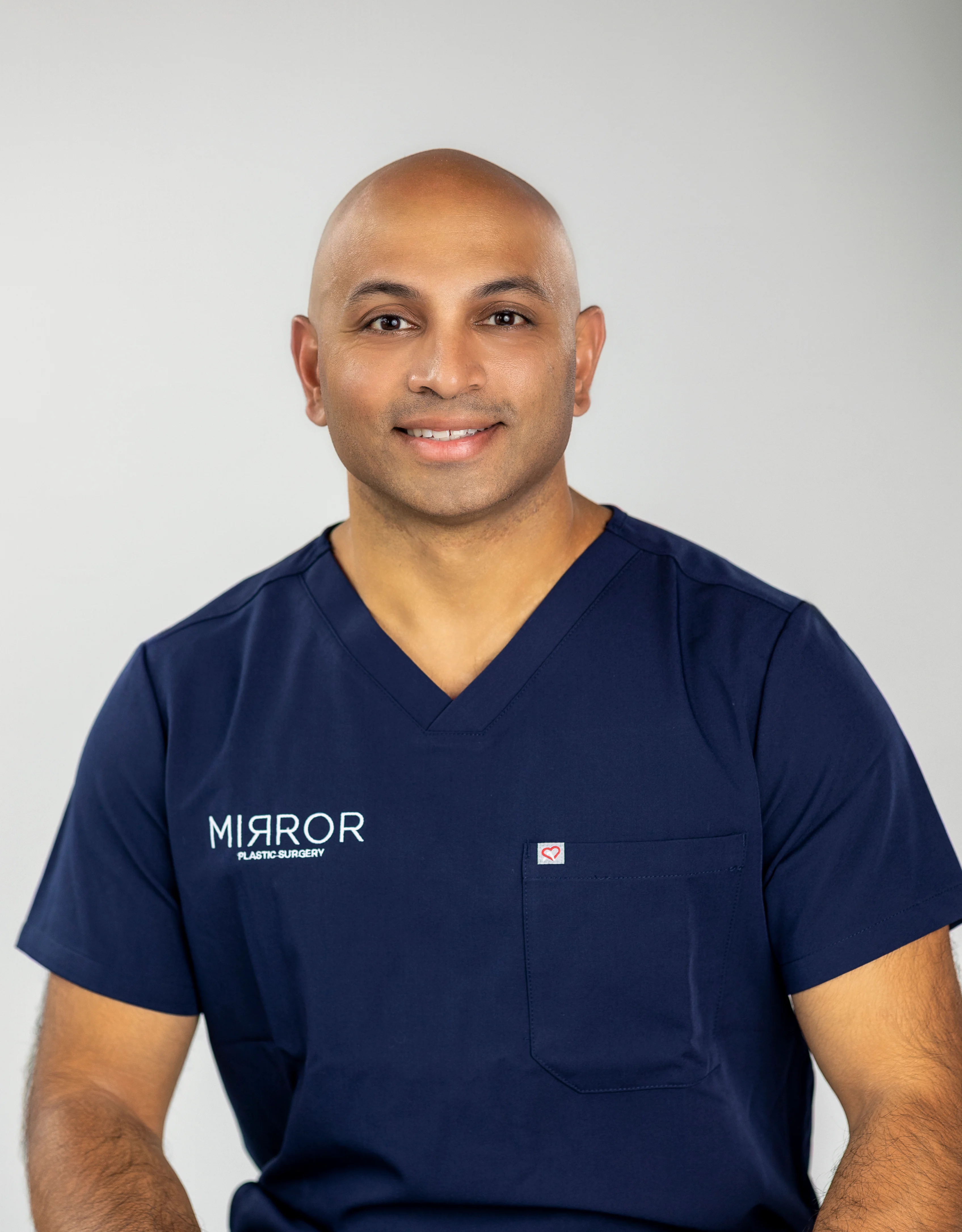
Learn how expert filler placement can support your natural movement and expression by booking a consultation with Dr. Akash at Mirror Plastic Surgery.
Disclaimer: Results may vary from person to person. Editorial content, before and after images, and patient testimonials do not constitute a guarantee of specific results.
1 Results may vary from person to person. Editorial content, before and after images, and patient testimonials do not constitute a guarantee of specific results.