

Written by: Ellie Pranckevicius, FNP-BC, Aesthetic Nurse Practitioner & Aesthetic Injector | Facial Restoration & Regenerative Injectable Specialist, Mirror Plastic Surgery | Last updated: August 6, 2026
Key Takeaways on Fillers and Facial Movement
- Repeated dermal filler sessions can restrict facial muscle movement over time by changing tissue glide planes and muscle leverage.
- Placement depth, G-prime selection, and cumulative volume in each facial zone largely determine whether filler supports or limits natural expression.
- High-volume or high-G-prime filler in mobile or avascular planes raises the risk of long-term stiffness and compensatory muscle patterns.
- Recovery of natural movement after filler dissolution usually follows stages, and complex cases may need 12–24 months for full baseline return.1
- Patients who want anatomy-first, movement-preserving filler treatment can schedule a personalized assessment with Ellie at Mirror Plastic Surgery.
How Fillers Restrict Muscle Movement Over Time
The relationship between injected filler and facial muscle function is not simply about how much product is used. Kapoor et al. (2021) in Clinical, Cosmetic and Investigational Dermatology showed that fillers placed within or immediately next to the SMAS and muscle layer can change muscle movement. The outcome depends on placement depth, product rheology, and cumulative volume, and these variables compound across repeated sessions as tissue glide planes gradually shift.
Filler persistence compounds the problem, especially in avascular planes where enzymatic degradation is limited. A 2026 histologically confirmed case report in Plastic and Reconstructive Surgery Global Open documented dermal filler migrating from a temporal injection site to both upper eyelids over two years, with basophilic dermal deposits dissociating collagen fibers. The authors identified the interfascial temporal space, an avascular gliding plane, as the key factor that allowed product to persist and mechanically interfere far longer than standard clinical timelines suggest.
Technique Checklist 1: Depth and Volume Fundamentals
- Confirm injection depth matches the tissue plane target: supraperiosteal for structural support, mid-dermis for fine lines, superficial fat for mobile zones.
- Limit per-site volume to 0.2–0.5 mL at deep structural points to avoid diffuse elevation across layers.
- Avoid SMAS-layer placement in high-movement zones where the risk of mechanical blocking is greatest.
- Select product G-prime appropriate to depth: high G-prime supraperiosteally, low G-prime in dynamic superficial zones.
- Document cumulative volume per anatomical zone across sessions, not just per visit.
Why Volume, Depth, and Layering Matter for Movement
Product rheology, specifically G-prime (elastic modulus), determines how a filler behaves under the mechanical load of muscle contraction. High-G-prime filler is reserved for skeletal support at the temple, zygomatic arch, nasal base, and chin. Low-G-prime product is better suited to dynamic zones such as nasolabial folds and marionette lines, where the goal is to preserve natural expression.
Kapoor et al. further explain that high-G-prime, high-cohesivity fillers placed supraperiosteally on bone create stable projection and lifting vectors. These vectors reposition retaining ligaments and indirectly influence muscle leverage without spreading under compression. Softer products in the same plane may instead migrate along tissue interfaces, which changes how forces travel through the face.
Ellie Pranckevicius’s conservative full-face layering protocol at Mirror Plastic Surgery applies these principles in daily practice. The protocol sequences treatment from deep structural foundations outward, which allows each layer to settle before the next is added. Within each layer, G-prime and cohesivity are matched to that tissue plane’s mechanical demands, such as high-modulus products on bone and low-modulus products in mobile zones. Only after this foundational restoration is complete does Ellie consider volume augmentation, which helps prevent the overcorrection that generates glide-plane restriction. A randomized comparator-controlled trial of high-G-prime chin fillers showed that precise product selection can reach the same aesthetic endpoints with less volume, supporting conservative-volume protocols as a structural advantage rather than a compromise.
Start with a personalized layering assessment to map your existing tissue planes and align product choice with your unique anatomy.
Technique Checklist 2: Layering Protocol Standards
- Sequence injections from deep to superficial within each session to avoid trapping product in intermediate planes.
- Use cannula delivery with low-to-medium cohesivity products in mobile zones, such as marionette lines and the perioral area, to support tissue integration.
- Reserve high-G-prime fillers for ligament-adjacent and supraperiosteal sites only.
- Apply low-modulus, tissue-adaptive fillers in superficial dynamic zones; low-modulus dermal filler gels placed per subunit have been shown to resorb over time with no delayed inflammatory reactions in prospective cohorts.
- Treat the full face as a single anatomical unit and avoid isolated single-area injections that create asymmetric mechanical loading.
How Nearby Muscles Compensate When Fillers Block Movement
When primary muscles of expression encounter mechanical resistance from filler deposits, adjacent and antagonist muscles recruit to maintain the same movement output. This over-recruitment often escapes early detection by patients and providers yet accumulates over repeated sessions. As the temporal migration case demonstrated, product displaced along glide planes under gravity and muscle action can reach anatomically remote sites, so the mechanical footprint of a single injection extends beyond the needle’s entry point.
Lymphatic clearance adds another layer of variability. Avascular planes with reduced lymphatic drainage, with the interfascial temporal space as a clear example, retain product longer and sustain mechanical interference with nearby muscle bellies across years rather than months. A 2026 narrative review by Alizadeh et al. confirms that dermal filler gels are biodegradable, yet clinical effects vary with product, technique, and patient-specific clearance factors. This range is wide enough to create significant differences between patients in how long compensatory patterns persist.
What Happens to Movement After Stopping Fillers
Recovery of natural movement after filler cessation or dissolution follows a staged timeline that depends on cumulative volume, product type, and placement depth. The table below maps this recovery across four phases, showing that superficial fillers may resolve within months, while deep or migrated product can require up to two years for full baseline return, which contrasts with the 12–18 month duration claims in most product labeling.1
| Time Post-Dissolution | Expected Movement Change | Supporting Evidence | Clinical Note |
|---|---|---|---|
| 0–4 weeks | Immediate reduction in bulk; some swelling may temporarily mask movement improvement | Alizadeh et al. 2026 | Dissolving agent breaks filler cross-links rapidly; multiple sessions may be required for high-volume or migrated product |
| 1–3 months | Glide-plane softening begins; early return of dynamic expression in superficially treated zones | JPRAS Open 2026 cohort | Low-modulus fillers in superficial planes resolve fastest; deep supraperiosteal deposits may persist longer |
| 6–12 months | Gradual softening and volume loss in mid-dermis and superficial fat; expression increasingly normalized | JPRAS Open 2026 cohort | Compensatory muscle recruitment patterns may require neuromuscular retraining in long-term patients |
| 18–24 months | Return to baseline movement in most treated sites for low-modulus products; higher-modulus or migrated product may need additional dissolution | JPRAS Open 2026 cohort | Histologically confirmed migration cases, such as temporal to periorbital, may require surgical removal if dissolving agent access is limited |
Injection Practices That Increase Stiffness Risk
Certain technique patterns consistently appear in the literature as predictors of long-term movement restriction. High-volume single-session loading, especially in deep fat or SMAS-adjacent planes, generates the diffuse multi-layer elevation that Kapoor et al. identify as the main mechanical pathway to glide-plane restriction. Single-plane injection strategies that ignore the deep-to-superficial architecture of facial tissue create uneven mechanical loading across muscle compartments.
Superficial placement of high-G-prime, high-cohesivity products in dynamic zones creates a separate but equally significant risk. Stiff gels in mobile tissue resist deformation under muscle contraction and behave like mechanical blocks described in the myomodulation literature. The 2026 temporal migration case also highlights fanning techniques in avascular planes as a pathway for remote migration, with product traveling along glide planes to distant sites over a two-year period.
Rushed, high-volume practices that ignore cumulative session volume per anatomical zone compound these risks with each additional treatment cycle. Patients who have received multiple sessions in such settings are the most likely to present with stiffness and expression restriction.
How to Choose an Injector Who Protects Natural Movement
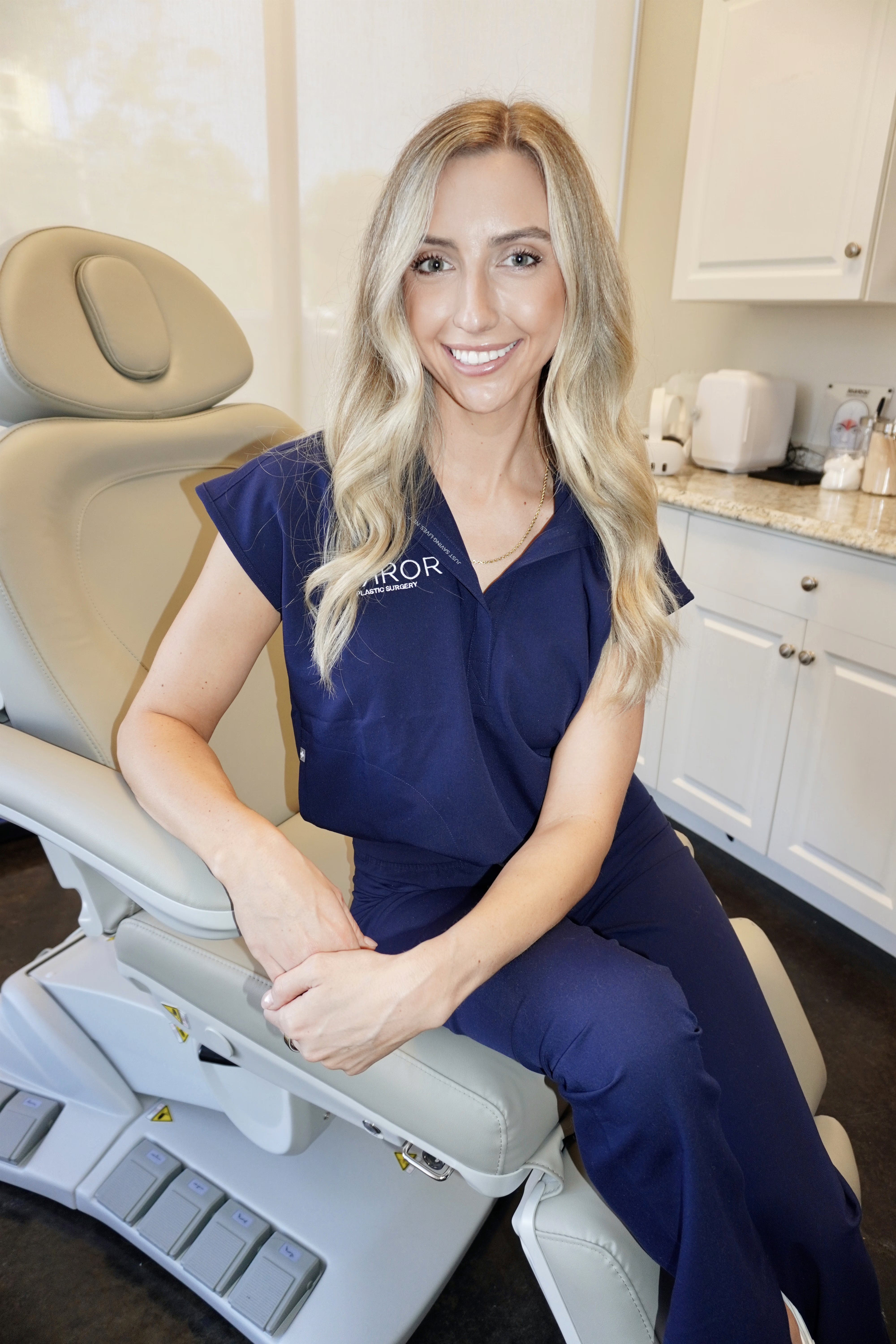
Choosing an anatomy-protective injector starts with clear, verifiable criteria. An injector should show formal training in subdermal and fascial anatomy, not just surface-level product knowledge. Ellie Pranckevicius, FNP-BC, has a dual background in esthetics and advanced nursing, including four years in the Neuroscience ICU at Tampa General Hospital, which supports a depth of physiologic understanding that is uncommon in aesthetic practice.
Conservative volume per session, combined with documented tracking of cumulative zone-specific volume across visits, forms a core safety standard. G-prime selection matched to tissue plane, full-face sequencing instead of isolated zone treatment, and honest communication about when additional volume is not appropriate work together as a single system. This combination protects natural movement while still addressing aesthetic goals.
Mirror Plastic Surgery’s one-hour top-to-bottom assessment provides the time and diagnostic structure to evaluate all of these variables for each patient’s anatomy. The practice’s concierge medicine model, which limits daily treatment volume to preserve focused clinical attention, contrasts with high-throughput environments where red-flag practices are more common.
Arrange a comprehensive, anatomy-first evaluation with Ellie at Mirror Plastic Surgery in St. Petersburg, Florida.
Frequently Asked Questions
Can fillers permanently restrict facial expressions?
Permanent restriction is not the usual outcome, yet prolonged mechanical interference is well documented. Dermal fillers placed in avascular gliding planes can persist for years beyond their labeled duration because reduced vascularity and lymphatic clearance limit enzymatic degradation. During that extended persistence, product can block muscle contraction amplitude or alter leverage vectors. Once product is fully dissolved or resorbed, a process that may take up to 24 months for some formulations and placements, movement generally returns toward baseline. Patients with many years of high-volume treatment may still have compensatory muscle recruitment patterns that outlast product clearance and benefit from targeted neuromuscular assessment.
What happens after 10 years of filler?
A decade of repeated filler sessions often leaves product across several tissue planes, especially in patients treated at high-volume practices without conservative volume management. Over that period, partially degraded or migrated filler fractions can dissociate collagen fibers in dermal and fascial planes, alter the mechanical environment of retaining ligaments, and shift leverage vectors of underlying muscles. The clinical picture often includes visible tissue heaviness, reduced expressivity, and asymmetry from uneven mechanical loading. Reversal remains possible but usually requires a staged dissolution plan and a full-face anatomical reassessment before any new product is added.
Does dissolving filler restore normal muscle movement?
Dissolution removes the mechanical obstruction created by the filler depot, which is the necessary first step toward movement restoration. In superficial and mid-dermal placements using low-modulus products, movement improvement can begin within weeks of dissolution.1 Deeper placements, migrated product, or high-cohesivity gels may require multiple dissolving agent sessions and a longer recovery window, and clinical evidence supports a 12-to-24-month timeline for full baseline return in the most affected sites.1 Dissolution does not automatically reverse compensatory muscle recruitment patterns that developed during the period of restriction, and those patterns may require separate assessment and management.
How do I know if my injector is using a safe technique?
A safe, anatomy-protective injector performs a thorough pre-treatment assessment that maps your tissue planes, documents prior treatment history including cumulative volumes, and selects product G-prime based on the depth and mobility of each target zone. They sequence injections from deep structural planes to superficial refinement within a single session, use conservative per-site volumes such as 0.2–0.5 mL at deep structural points, and treat the face as an integrated anatomical unit rather than isolated problem areas. Clear boundaries around when additional volume is not appropriate, even when requested, signal an injector who prioritizes function and long-term anatomy over short-term volume gains.
Conclusion: Protecting Expression While Using Fillers
The long-term effects of facial dermal fillers on muscle movement are real, documented, and strongly influenced by technique. Placement depth, G-prime selection, per-session volume, and cumulative loading in each zone determine whether filler supports or restricts natural expression over time. Conservative, layered, anatomy-first injection protocols, grounded in the evidence reviewed here, represent the standard of care for patients who value function alongside aesthetics.
Mirror Plastic Surgery’s approach, led by Ellie Pranckevicius within the clinical framework established by Harvard-educated, Johns Hopkins-trained plastic surgeon Dr. Akash Chandawarkar, follows these priorities in sequence: safety first, function second, aesthetics third.
Begin your one-hour assessment at Mirror Plastic Surgery’s St. Petersburg practice to receive a comprehensive evaluation of your facial anatomy and treatment history.
Disclaimer: Results may vary from person to person. Editorial content, before and after images, and patient testimonials do not constitute a guarantee of specific results.
References
- El Feghaly C, Tawa P. Chronic upper eyelid edema following temporal hyaluronic acid filler injection: a histologically confirmed case of remote migration. Plastic and Reconstructive Surgery Global Open. 2026.
- Alizadeh et al. Narrative review of hyaluronic acid filler biodegradability and clinical duration. Plastic and Reconstructive Surgery Global Open. 2026.
- Kapoor et al. Tissue plane mechanics and myomodulation in hyaluronic acid filler placement. Clinical, Cosmetic and Investigational Dermatology. 2021.
- Wu et al. The L-V approach to facial rejuvenation: ligament-targeted and plane-sequenced filler protocol. Plastic and Reconstructive Surgery Global Open. 2026.
- 24-month prospective cohort of low-modulus hyaluronic acid filler (Volonic Light). JPRAS Open. 2026.
- Randomized comparator-controlled trial of high-G-prime chin fillers: volume efficiency and aesthetic outcomes. Aesthetic Surgery Journal. 2024.
- Prospective clinical trial of novel crosslinked hyaluronic acid filler: 9-month outcomes in 203 participants. Aesthetic Surgery Journal Open Forum. 2026.
1 Results may vary from person to person. Editorial content, before and after images, and patient testimonials do not constitute a guarantee of specific results.