

Written by: Dr. Akash Chandawarkar, Board Certified Plastic Surgeon, Mirror Plastic Surgery | Last updated: September 9, 2026
Introduction
Facial fillers can restore youthful volume when they respect the face’s layered anatomy.1 Natural results depend on precise placement, conservative volume, and a provider with deep anatomical training.1 This article explains how anatomy guides safe, subtle filler treatments and why your choice of injector matters more than the brand of filler.
Why Facial Anatomy Is The Key To Natural Results
The “pillow face” look, with overinflated cheeks and distorted lips, usually reflects technique and planning rather than a problem with filler products. A 2026 narrative review in Plastic and Reconstructive Surgery Global Open links facial overfilled syndrome to inadequate anatomical knowledge, inaccurate pretreatment assessment, improper injection technique, inappropriate product choice, and overfilling.
Natural volume restoration requires understanding where volume has been lost and replacing it in the specific anatomical layers where it originally existed. A board-certified plastic surgeon views the face as a three-dimensional structure with distinct layers: bone, fat compartments, retaining ligaments, and muscle. Each layer ages differently and calls for a tailored treatment strategy.
The Aging Face: Volume Loss Layer By Layer
Think of the face as a tent. The bones act as poles, the retaining ligaments function as ropes, and the fat compartments form the fabric. As the poles shrink and the ropes loosen, the fabric sags and loses support. Facial aging involves changes in bone, fat, and ligaments, not just wrinkles.
Bone: The Shrinking Foundation
The facial skeleton changes over time. Bone resorption occurs in key areas such as the orbital rim, maxilla (midface), and mandible (jawline). Aging involves three-dimensional structural changes including skeletal resorption in the maxilla, mandible, and orbital apertures, which explains why structural fillers for chin and jawline contouring can restore youthful proportions1. As these bones lose volume, the soft tissues above them lose their foundation.
Fat Compartments: The Deflating Volume
The face contains distinct superficial and deep fat compartments that age at different rates. A 2026 cross-sectional MRI study of 47 healthy female volunteers aged 18–65 found heterogeneous age-related changes in facial fat distribution, with buccal fat pad and nasolabial fold volumes decreasing with age and infraorbital fat volume increasing significantly.
The deep medial cheek fat (DMCF), located over the maxilla, contributes heavily to anterior projection and deflates significantly with age. At the same time, ultrasound-based research by Professor Sebastian Cotofana shows that superficial fat compartments are highly mobile, while deep compartments remain stable. Deep compartments therefore make preferred targets for filler placement, because augmenting deep volume can reposition superficial fat compartments.
Retaining Ligaments: The Loosening Support
The zygomatic, masseteric, and mandibular ligaments anchor facial soft tissues to the underlying skeleton. A 2026 review in Clinical, Cosmetic and Investigational Dermatology identifies elongation and loss of tensile support of retaining ligaments as key pathological factors in facial aging, with reduced tensile integrity contributing directly to inferior displacement of soft tissues and contour breakdown. Nasolabial folds deepen and jowls form because this structural support weakens beneath the skin.
The Layered Anatomy: A Surgeon’s Perspective On Filler Placement
The face is organized into five layers: skin, superficial or subdermal fatty layer, superficial fascia including muscles, deep fatty layer, and periosteum. A skilled injector selects the correct layer for each aesthetic goal. Broadly, filler placement falls into two categories: deep placement for structural support and superficial placement for contour refinement.
Deep Placement: Structural Support
Fillers placed deep on the bone (supraperiosteal) or within deep fat compartments provide structural support and lift. The dual-plane approach, systematically described by Italian surgeon Giovanni Salti, differentiates filler roles by depth. High-elasticity (high G′) firm fillers in the deep supraperiosteal or sub-SMAS plane provide structural support against gravitational descent. High-elasticity fillers work well in this plane because they hold their shape against gravitational forces.
Superficial Placement: Contour Refinement
Low-elasticity, highly cohesive fillers in the superficial subdermal plane transmit lifting effects and absorb shear stress for natural contour refinement. Overfilled results often appear when too much filler sits superficially without first addressing deeper structural layers. That mismatch between product, plane, and purpose creates a puffy, unnatural look.
Five Facial Areas Where Anatomy Matters Most
Every facial region has its own anatomical nuances, yet five areas especially highlight how anatomy shapes natural results. Each of these regions requires a tailored plan for volume restoration and contour.
1. Midface (Cheeks)
2. Temples
Temporal hollowing develops as fat and muscle atrophy with age. Injection in this region must respect the deep fascial layers and protect the temporal artery branches. Proper temple volume restoration supports the brow and upper face and creates a smooth transition between facial regions.
3. Tear Trough
This delicate area demands precise placement to avoid lumps, the Tyndall effect (bluish discoloration from superficial filler), and chronic swelling. A 2026 MRI study found filler in the tear trough region expanded to a mean thickness of 7.17 mm, the greatest proportional increase of any region studied, likely due to dense orbicularis retaining ligaments and limited subcutaneous space.
4. Jawline
The mandibular ligaments anchor the tissue of the lower jaw, and their laxity contributes to pre-jowl hollowing and loss of crisp jawline definition. Injecting along the mandible can define the jawline and address pre-jowl hollowing. Accurate knowledge of the mandibular ligament’s location is critical, because injection in the wrong plane can accentuate jowls instead of improving them.
5. Lips
Respecting the vermilion border and understanding the mucosal layer’s unique anatomy helps prevent a “duck lip” appearance. Lip injections require a different strategy than structural facial fillers, with more superficial placement and softer products that match the tissue’s natural flexibility.
These anatomical zones form the foundation of natural results. A board-certified plastic surgeon with advanced aesthetic training applies this knowledge to every injection. Schedule a personalized facial anatomy consultation to review your features and goals with Dr. Akash Chandawarkar.
Safety First: Danger Zones And How Anatomy Prevents Complications
Certain facial regions carry higher risks of vascular complications because of their arterial anatomy. A 2026 systematic review in Aesthetic Plastic Surgery found that the glabella and nasal dorsum were most frequently implicated in blindness, while the nasolabial fold and forehead were associated with cerebral embolic events, and concluded that anatomic location outweighs injected volume in predicting risk.
Key danger zones include the glabella, nasal dorsum, tear trough, and temporal region. The glabella carries a high risk of vascular occlusion because of the supratrochlear and supraorbital arteries. The nasal dorsum connects with the ophthalmic artery and can allow retrograde embolism. The tear trough can develop the Tyndall effect and vascular compromise if injected too superficially. The temporal region contains deep temporal artery branches that require precise anatomical awareness.
Intra-arterial injection with subsequent necrosis occurs in 0.1–0.3% of filler procedures, and blindness represents the most devastating and consistently irreversible complication. This pattern underscores the importance of choosing a board-certified plastic surgeon. Dr. Akash Chandawarkar’s surgical background provides a three-dimensional understanding of facial vascular anatomy that many non-physician injectors lack. Board-certified dermatologists or plastic surgeons are more likely to achieve sophisticated outcomes because their residency training covers facial anatomy, vascular networks, and emergency complication management1.
Safety and natural results align when your injector has surgical-level anatomical training. Discuss your safety-focused filler plan and learn how Dr. Akash prioritizes risk reduction at every step.
Do Fillers Age You Over Time? What The Research Says
A 2026 MRI study found that the volume of filler detected on MRI averaged 2.8 times the originally injected volume. Detectable filler persisted for up to 15 years after injection, with expansion driven by the filler’s hydrophilic properties drawing in water. The authors recommend a conservative approach to injection volumes to avoid overcorrection.
A separate 2026 review concludes that fillers are biodegradable and temporary, with clinical effects generally lasting 4–18 months depending on product, technique, and patient factors. The review notes that the risk of gel migration or facial overfill syndrome relates to inadequate anatomical knowledge, suboptimal technique, or poor patient selection rather than an inherent flaw in the products.
The concept of “filler fatigue,” where repeated injections supposedly stretch the skin, has not been proven in controlled studies. Research does show that chronic overfilling can stretch tissues over time. Strategic, conservative treatment with thoughtful maintenance helps preserve tissue quality.
Which Filler Looks Most Natural? Product Selection In Context
Natural-looking results depend more on injection technique and anatomical placement than on brand names.1 An international consensus paper in the Journal of Cosmetic Dermatology proposes the Natural Outcomes Framework, defining natural outcomes across four dimensions: see (visual observation), touch (palpation), feel (patient satisfaction), and express (dynamic facial movement). The authors emphasize that technique, anatomy knowledge, and understanding of aging are as important as product choice.
A filler’s firmness, measured by its elastic modulus (G-prime), guides ideal placement. High G-prime gels work best deep over bone for structural support in areas like the chin and cheeks, while low G-prime gels suit lips and fine lines. The most appropriate filler mimics the natural tissue in the target layer, and the injector must know how to place it accurately.
Dr. Akash Chandawarkar serves on advisory boards for Merz Aesthetics (Radiesse) and Tiger Aesthetics (AlloClae), which gives him insight into product development that many injectors do not have. This supplier-neutral perspective allows him to recommend products based on your anatomy and goals rather than on commission structures.
Choosing Your Injector: Why Anatomical Expertise Matters
Current research suggests clear criteria for selecting a provider for dermal filler treatments. Board certification in plastic surgery or dermatology indicates residency training that includes comprehensive facial anatomy education. Specialized fellowship training in aesthetic surgery and facial anatomy builds on that foundation. Experience managing complications shows that a provider understands vascular emergencies and how to avoid them. A treatment philosophy that prioritizes safety, function, and natural results over volume supports long-term facial health.
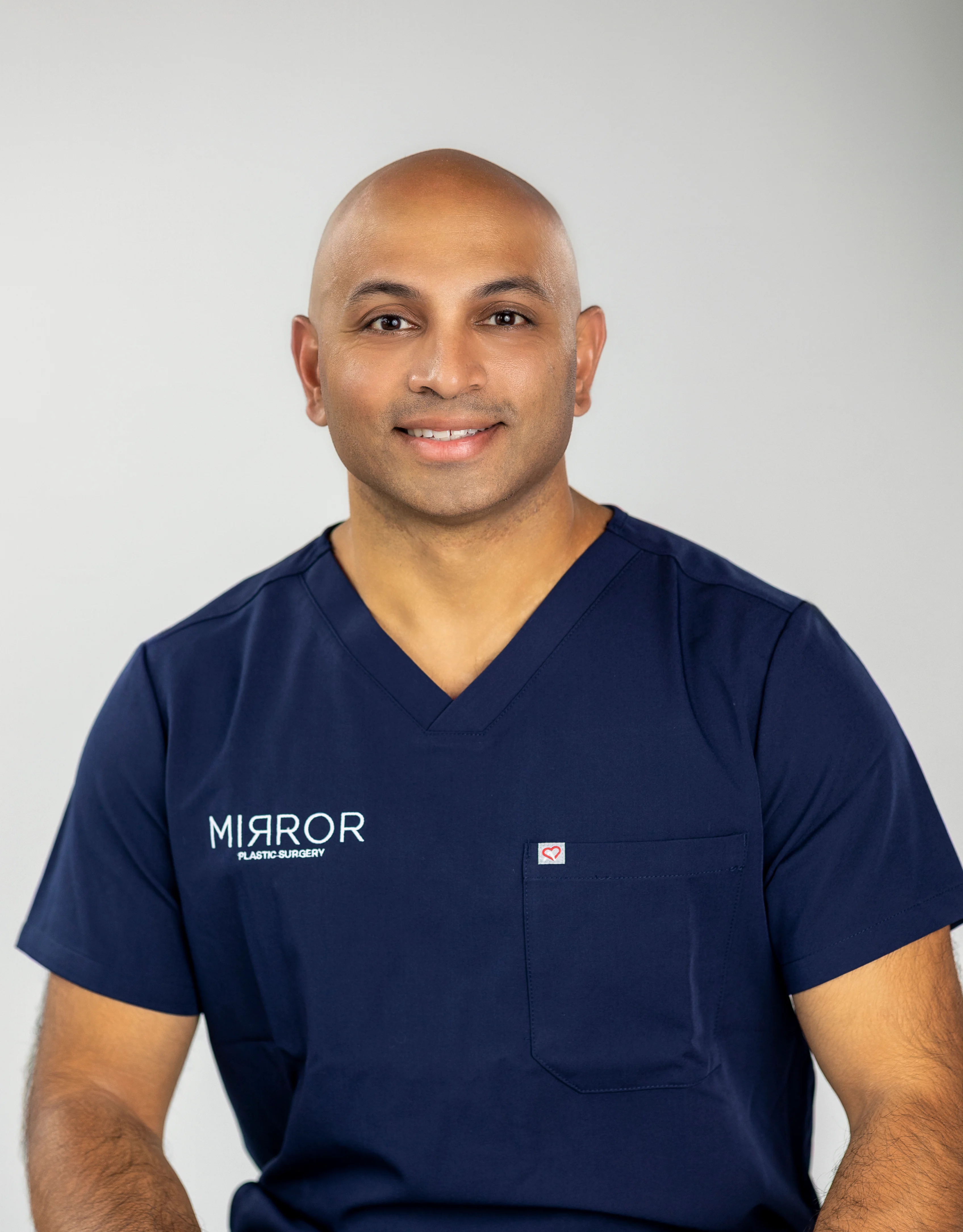
Dr. Akash Chandawarkar meets these standards. He is a Harvard-educated physician, Johns Hopkins-trained plastic surgeon, and board-certified by the American Board of Plastic Surgery. He completed fellowship training in aesthetic surgery at the Manhattan Eye Ear & Throat Hospital (MEETH)/Lenox Hill Hospital. At Mirror Plastic Surgery in St. Petersburg, FL, Dr. Akash personally performs all injectable treatments, so patients work directly with the surgeon throughout their care.
Conclusion
Facial aging involves bone resorption, fat compartment deflation, and retaining ligament laxity, along with surface wrinkles. Fillers work best when placed in specific anatomical layers, with deep injections providing structural support and superficial injections refining contour. Natural results come from a holistic, full-face approach guided by anatomy, conservative volume, and respect for danger zones.1
Your choice of provider influences outcomes more than the specific filler brand. A surgeon with advanced anatomical training can restore volume while preserving your unique expression. Schedule your personalized consultation to receive a top-to-bottom assessment and a tailored treatment plan at Mirror Plastic Surgery in St. Petersburg, FL.
Disclaimer: Results may vary from person to person. Editorial content, before and after images, and patient testimonials do not constitute a guarantee of specific results.
1 Results may vary from person to person. Editorial content, before and after images, and patient testimonials do not constitute a guarantee of specific results.