
Written by: Dr. Akash Chandawarkar, Board Certified Plastic Surgeon, Mirror Plastic Surgery | Last updated: September 9, 2026
Key Takeaways
- BBL fat necrosis happens when some transferred fat cells lose blood supply and die, creating firm lumps that usually fade over time.
- Symptoms often appear 2 to 6 weeks after surgery and include hard nodules, tenderness, discoloration, and contour changes, though most cases remain benign.1
- Most fat necrosis lumps improve within 4 to 12 weeks, with full resolution in 3 to 6 months, although larger areas or risk factors can slow healing.1
- Gentle lymphatic massage may help soften lumps when your surgeon approves it, while forceful massage can damage the graft and worsen symptoms.
- Persistent lumps or worrisome symptoms should prompt a consultation at Mirror Plastic Surgery for expert evaluation and tailored treatment.
What Fat Necrosis Means After A BBL
During a Brazilian Butt Lift, your surgeon harvests fat from donor areas and injects it into the buttocks in precise layers. For these transferred fat cells to survive, they must connect to a new blood supply within the first few days. Cells more than roughly two millimeters from a blood supply rarely survive, which explains why fat necrosis develops.
The process unfolds in stages. Ischemia causes fat cell death, then triglycerides break down into free fatty acids that bind with calcium in a process called saponification. The body responds with inflammation. Macrophages and neutrophils move in, foam cells and multinucleated giant cells form, and fibrosis and encapsulation create the firm nodules you can feel.
Not all firmness after a BBL represents fat necrosis. Some tightness and firmness are normal parts of healing and tissue remodeling. True fat necrosis lumps usually feel firmer, more defined, and last longer than general post-surgical firmness.
Common Signs And Symptoms After BBL
Fat necrosis usually becomes noticeable two to six weeks after surgery, once early swelling settles and the underlying tissue becomes easier to feel.1 Typical symptoms include:
- Hard, firm lumps or nodules beneath the skin
- Tenderness or mild pain where the lump sits
- Skin discoloration, such as lingering bruising or redness
- Irregular contour or visible asymmetry
- A gravel-like or pebbled texture in some areas
- Oil cysts, which are smooth, fluid-filled pockets formed when released triglycerides pool and surrounding tissue forms a capsule
These lumps usually stay benign, yet they still deserve attention. Concerning signs include firm, immobile lumps that do not soften, localized warmth, redness, tenderness, skin dimpling, or drainage of thin oily fluid.
If you feel unsure about a lump or change, book a consultation with Ellie at Mirror Plastic Surgery for a personalized assessment.
Typical Timeline For Fat Necrosis After BBL
Most fat necrosis lumps start to soften within 4 to 12 weeks as swelling decreases and tissues relax, with some areas taking 3 to 6 months to fully resolve.1 In some BBL cases, small lumps can linger for a year or longer while the body slowly breaks down and absorbs dead fat cells.1
Several factors shape how long fat necrosis lasts:
- Size Of The Affected Area, because larger areas take longer to clear and sometimes become infected
- Smoking Status, since smoking reduces blood flow and clearly lowers fat graft survival
- BMI And Metabolic Health, as 50% of patients with BMI above 30 kg/m² developed fat necrosis compared with none in the non-obese group in one series
- Pressure Compliance, because direct pressure or shear forces in early healing can kill transferred fat cells and worsen necrosis
- Surgical Technique, since large single boluses create more necrosis than the same volume placed in thin, separated strands across several planes
Most cases improve without medical procedures. Lumps that last beyond six months or cause ongoing discomfort should be evaluated, since targeted treatments can speed resolution.
Safe Use Of Massage For BBL Lumps
Gentle massage can sometimes soften lumps and support circulation, yet it does not guarantee complete resolution. Aggressive massage can injure the graft and worsen contour problems. Massage directly on fat implantation sites is specifically contraindicated early in recovery because pressure can shift fat, causing disfigurement, pain, swelling, or circulation issues.
With surgeon approval, gentle lymphatic massage may begin as early as 24 to 72 hours after surgery. Deeper massage for lumps usually starts around two weeks post-op, although direct massage on the buttocks remains discouraged because it can damage transferred fat cells.
Lymphatic drainage massage helps reduce swelling and support circulation, which may soften lumps and speed recovery. A qualified therapist should use light, superficial strokes that avoid compressing the graft. Always clear any massage plan with your surgeon first.
Post-Operative BBL Smell And When To Worry
A mild, musty odor during the first 7 to 10 days after a BBL is common and usually comes from trapped tumescent fluid, old blood, and garment buildup. Good hygiene and garment care usually resolve this quickly.
A strong, foul, or persistent odor from an incision site signals a problem. Possible causes include:
- Infection that needs medical treatment
- An infected oil cyst
- Necrotic tissue that requires professional care
A strong, foul, pungent, or sweet rot-like odor from incision sites, especially with fever above 38.5°C (101.3°F), spreading redness, warmth, or thick pus, may indicate infection and needs urgent medical evaluation. Most fat necrosis cases do not involve odor, yet odor with these symptoms requires immediate attention.
Home Care Tips For BBL Fat Necrosis
Several evidence-based steps at home can support your body as it heals:
- Wear Compression Garments As Directed, since they support healing and help control swelling
- Stay Hydrated, because good hydration supports tissue health and circulation
- Eat A Nutrient-Rich Diet, with vitamins A and C to support tissue repair
- Avoid Smoking And Alcohol, as smoking and vaping restrict blood flow, impair new vessel formation, and raise the risk of delayed healing and fat loss
- Apply Warm Compresses only with surgeon approval, and remember that ice packs should never go on the buttocks after a BBL because cold-induced vasoconstriction can kill the graft
- Take Over-The-Counter Anti-Inflammatory Medication when appropriate, since NSAIDs for one to two weeks can ease symptoms during the acute phase if they are safe for you
Avoid draining, squeezing, or puncturing any lump yourself. Puncturing an oil cyst wall can worsen inflammation and should be avoided, especially during the acute phase. These home steps support healing but never replace professional evaluation.
Medical Treatments For Persistent Lumps
When lumps last beyond six months or cause pain or contour issues, a board-certified plastic surgeon may suggest one or more of these options:
- Steroid Injections to calm inflammation and speed softening
- VASER Liposuction Or Radiofrequency-Assisted Liposuction to emulsify necrotic fat and soften fibrotic tissue in moderate cases
- Needle Aspiration for liquid oil cysts, which can be quick and sometimes curative, although recurrence can occur
- Surgical Excision for large, painful, or cosmetically significant solid masses that do not respond to less invasive treatments
At Mirror Plastic Surgery, Dr. Akash Chandawarkar provides detailed evaluations and evidence-based treatment plans for patients with persistent fat necrosis. Book a consultation with Ellie to review your case with a board-certified plastic surgeon.
When To Contact Your Surgeon
Most fat necrosis cases improve on their own. Certain symptoms, however, require immediate medical attention:
- Severe Pain Or Swelling that worsens instead of improving after the first two weeks
- Redness, Warmth, Or Fever, especially fever above 38.5°C (101.3°F) or visible wound changes
- Foul-Smelling Drainage from any incision or lump
- Lumps That Grow Quickly or change in texture
- Worsening Asymmetry or contour irregularities that fail to improve
- Sudden Shortness Of Breath Or Chest Pain, which can signal fat embolism or pulmonary embolism and needs emergency care
A localized hard lump that feels different from general swelling should always prompt a surgeon visit. Many findings turn out to be normal healing, and professional reassurance offers real peace of mind.
Why Patients Choose Mirror Plastic Surgery
Mirror Plastic Surgery serves St. Petersburg and Tampa as a focused practice for BBL care and complication management. The practice is led by Dr. Akash Chandawarkar, a Harvard-educated, Johns Hopkins-trained plastic surgeon with advanced aesthetic fellowship training at Manhattan Eye Ear & Throat Hospital and Lenox Hill Hospital.
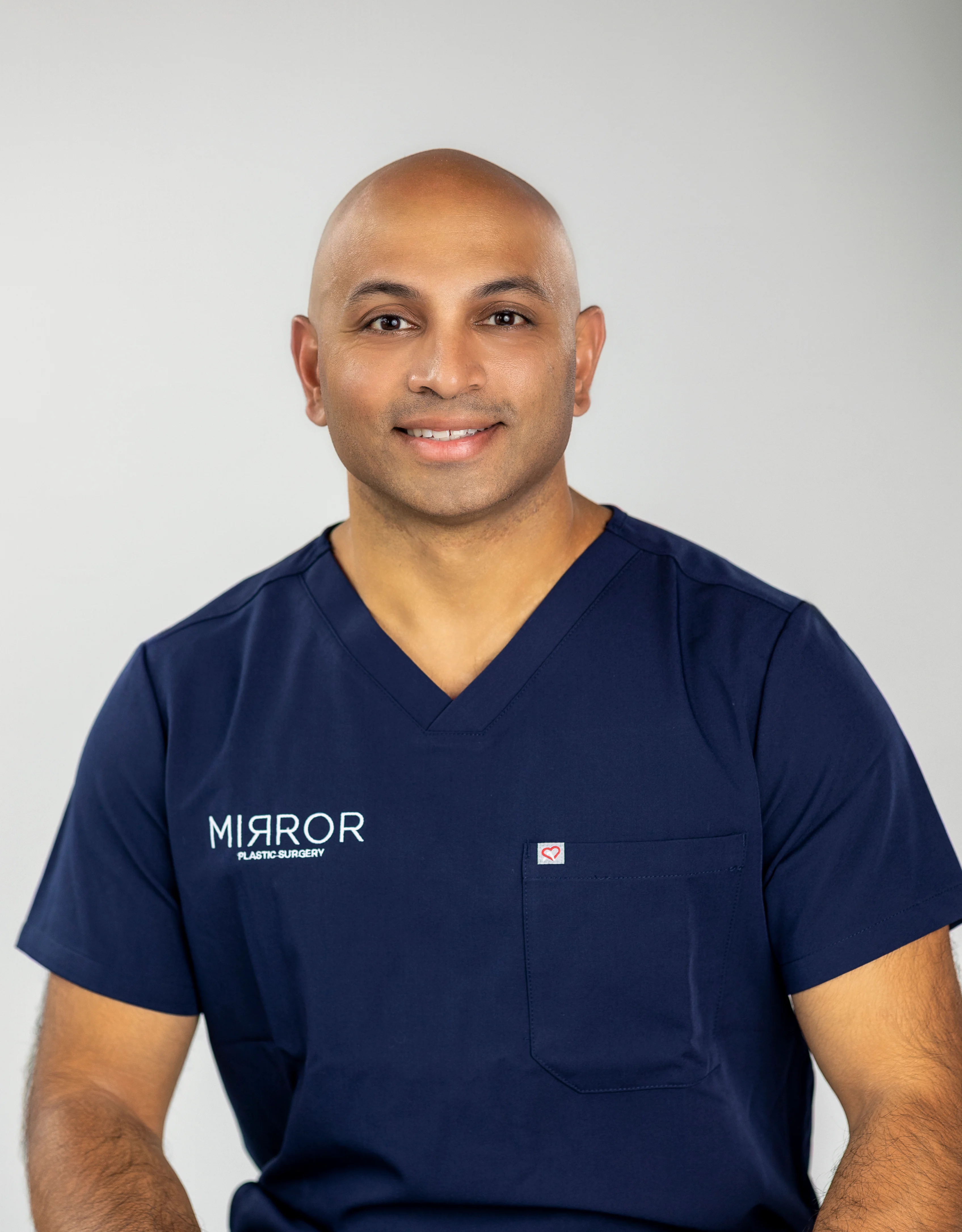
Consultations at Mirror Plastic Surgery can last up to an hour so your concerns receive full attention before any plan is made. The team schedules only one to two surgeries per day, which keeps the focus on you before, during, and after your procedure. Dr. Akash follows a safety-first philosophy that places function and health at the center of every decision.
Patients who feel worried about fat necrosis after a BBL elsewhere, or who plan a future BBL and want a clear understanding of risks, receive honest, personalized guidance. Book a consultation with Ellie and speak with a board-certified plastic surgeon who takes time to listen.
Frequently Asked Questions About BBL Fat Necrosis
Is Fat Necrosis After BBL Dangerous?
Fat necrosis itself is benign and reflects your body’s normal response to fat cells that did not survive transfer. The body walls off dead tissue and forms firm nodules or oil cysts. Most cases resolve without treatment. Fat necrosis differs from fat embolism, a more serious complication linked to improper injection technique during surgery rather than the healing phase.
Can Fat Necrosis Turn Into Cancer?
Fat necrosis does not turn into cancer. Fat necrosis is not precancerous and does not raise cancer risk. It can, however, mimic cancer on imaging, especially mammograms, because late-stage calcifications can resemble malignant findings. If you had a BBL and later undergo breast or body imaging, tell your radiologist about your surgery so they can interpret results accurately and avoid unnecessary follow-up.
Will Fat Necrosis Change My Final BBL Results?
Minor fat necrosis usually does not change your final shape in a noticeable way.1 The body gradually reabsorbs dead tissue, and surviving fat continues to integrate and maintain volume. Larger areas of necrosis can create contour irregularities or asymmetry.1 Surgeons typically assess and correct these only after full healing, usually between six and twelve months post-surgery.
Can I Lower My Risk Of Fat Necrosis?
You can significantly lower your risk with the right choices. Selecting a board-certified plastic surgeon who uses subcutaneous-only injection and moderate fat volumes per pass plays the biggest role. On your side, avoid sitting directly on your buttocks for the period your surgeon recommends, usually four to six weeks. Wear your compression garment as directed, stop smoking well before surgery and throughout recovery, and keep your weight stable. These habits protect the fragile capillary networks that nourish new fat cells.
How Soon After BBL Can I Massage Lumps?
Start massage only after your surgeon gives clear approval. Many surgeons recommend gentle lymphatic drainage massage within the first week after surgery, sometimes as early as 24 to 72 hours, once they confirm that you are ready. Massage that starts too early or uses too much pressure can displace fat, disrupt healing tissue, and worsen lumps. Lymphatic drainage massage with light, superficial strokes by a trained therapist remains the safest option and should always be coordinated with your surgical team.
Your Next Step Toward Confidence
BBL fat necrosis occurs frequently, usually stays harmless, and often fades with time. Clear information about what you feel, what helps, and what requires attention can replace fear with confidence. Home care supports recovery, while professional guidance protects your safety and results.
At Mirror Plastic Surgery, Dr. Akash Chandawarkar combines board-certified expertise with a concierge-style experience and direct, honest communication. Your health and confidence guide every recommendation.
Call 727-361-6515 or text the office to reach the team. To schedule online, book a consultation with Ellie today.
Disclaimer: Results may vary from person to person. Editorial content, before and after images, and patient testimonials do not constitute a guarantee of specific results.
1 Results may vary from person to person. Editorial content, before and after images, and patient testimonials do not constitute a guarantee of specific results.