

Written by: Dr. Akash Chandawarkar, Board Certified Plastic Surgeon, Mirror Plastic Surgery | Last updated: September 9, 2026
Key Takeaways
- Granulomas and nodules after facial dermal fillers are uncommon, with low reported incidence for hyaluronic acid fillers and even lower rates for true granulomas.
- Most early lumps after filler represent product pooling or minor irregularities, while true granulomas usually develop months to years later.
- Delayed nodules and granulomas can involve biofilms, immune responses to illness or vaccination, and filler properties such as cross-link density.
- Accurate diagnosis relies on high-frequency ultrasound and evaluation by a board-certified plastic surgeon, and most nodules improve with conservative or targeted treatment.
How Common Are Granulomas And Nodules After Fillers?
Granulomas and nodules after dermal fillers occur infrequently, and rates vary by filler type and study design. Reported ranges include 0.02%–0.4% for hyaluronic acid and 0.04%–0.3% for bovine collagen, with some broader estimates extending up to 2.8% of treatments. For hyaluronic acid (HA) fillers, which are the most widely used, histologically confirmed foreign body granulomas fall within the same low range for delayed nodules overall, based on a 2026 narrative review in the Journal of Cosmetic Dermatology.
A 2022 retrospective study of 2,139 HA filler patients reported delayed nodules in 0.33% of cases, and only one case met biopsy criteria for a true foreign body granuloma. These findings support the conclusion that true granulomas are rare events.
Most post-filler lumps do not represent granulomas. They often reflect localized product pooling, minor technique-related irregularities, or short-lived swelling. The American Academy of Ophthalmology reports that more than 90% of HA filler adverse events are mild and transient, such as temporary redness, swelling, or bruising at the injection site.
Key Statistics
- Reported granuloma incidence varies by filler type and study, with some ranges extending up to 2.8% of treatments.
- In a 2,139-patient HA filler study, delayed nodules occurred in 0.33% of patients.
- EyeWiki clinical reference cites a delayed nodule incidence of 0.5% for HA fillers.
- More than 90% of HA filler adverse events are mild and short-lived.
Incidence By Filler Type: How Different Products Compare
Different filler categories carry different nodule and granuloma risks. The table below summarizes key data from peer-reviewed sources for the most studied filler types.
Low overall incidence of delayed nodules4 weeks to over 1 yearmost adverse events are mild and need no treatment1–3 cases per controlled facial injection trialAround 7 months medianNo enzymatic reversal; management focuses on prevention and supportive care1.0% in a 2023 FDA trial (9 mL dilution) vs. 17.2% in a 2009 trial (5 mL dilution)9–14 months mean; some cases beyond 24 monthsRates dropped sharply with higher dilution volumes and updated protocols
| Filler Type | Reported Incidence of Nodules/Granulomas | Typical Onset | Notes |
|---|---|---|---|
| Hyaluronic Acid (HA) | |||
| Calcium Hydroxylapatite (CaHA) | |||
| Poly-L-Lactic Acid (PLLA) |
The PLLA data highlight how technique changes can reshape risk. A chart review of 150 patients showed that a 10–12 mL dilution reduced nodule rates to 1.3% compared with 8% using a standard protocol. This finding shows that protocol refinement can significantly improve patient safety.
Understanding when these complications appear helps patients and providers respond quickly. The next section outlines typical timelines for early and delayed reactions.
Timeline For Filler Nodules And Granulomas
Most early nodules, felt within days to about two weeks after injection, represent non-inflammatory product pooling rather than granulomas. Lumps that appear immediately to within 2 weeks are usually non-inflammatory, while red and painful reactions beyond 2 weeks, especially around 3 to 4 months, reflect a different delayed pattern.
Delayed nodules show a broader and more variable timeline. A 2024 retrospective study in Frontiers in Microbiology reviewed 61 patients with delayed or late complications and found an average onset of 16.2 months, ranging from 3 months to 6 years. A 2025 systematic review in the Journal of Cosmetic Dermatology reported detection times from 1 week to 15 years, with a median of about 20 months.
Onset also depends on filler composition. The same 2025 review reported median granuloma onset of about 7 months for CaHA, 13 months for polycaprolactone (PCL), 19 months for PLLA, and up to 35 months for PMMA.
Why Delayed Nodules And Granulomas Develop
Current evidence points to three overlapping mechanisms that can act alone or together in a single lesion.
- Biofilms: Bacterial communities can attach to filler material and remain dormant until a systemic trigger occurs. A 2009 study identified bacteria with Gram stain and fluorescence in situ hybridization in tissue from 7 of 8 patients with long-lasting nodules despite negative cultures, showing that standard cultures may miss biofilm-related infection.
- Immune Triggers: Viral illnesses, including COVID-19, dental work, and vaccinations can provoke delayed immune reactions around existing filler. Up to 71% of delayed reactions occurred between September and December, overlapping with peak viral infection season.
- Product Properties: Higher cross-link density and chemical modification in HA fillers increase resistance to breakdown but also extend exposure to modified HA and residual cross-linker. This extended exposure may raise the likelihood of delayed inflammatory reactions or foreign body granulomas.
How Providers Diagnose Lumps And Nodules
Diagnosis starts with a focused physical exam and often includes high-frequency ultrasound. Ultrasound with 15–22 MHz probes is highly sensitive for detecting filler-related lesions. Imaging patterns differ by tissue type: filler deposits appear as well-defined anechoic pockets, while granulomas and fibrosis show more heterogeneous, ill-defined signals. Biopsy remains uncommon and is reserved for unclear or atypical cases.
Accurate interpretation requires deep anatomical knowledge. A board-certified plastic surgeon understands facial tissue planes, fascial layers, and vascular anatomy at a surgical level, which supports precise diagnosis and safe management.
Evidence-Based Treatment Options For Filler Nodules
Treatment plans depend on filler type, nodule characteristics, and whether infection or significant inflammation is present.
For HA Fillers
- Hyaluronidase injected directly into the nodule is the first-line approach. Longstanding lesions may need several sessions because fibrotic tissue can partially encapsulate filler.
- Ultrasound-guided injections allow direct visualization of filler deposits, improving enzyme placement, local concentration, and contact with the target area.
- Intralesional corticosteroids such as triamcinolone acetonide often accompany hyaluronidase. Repeated low-dose injections spaced over weeks help calm the immune response and soften fibrotic capsules.
For Inflammatory Or Infectious Nodules
- Oral antibiotics, including macrolides and tetracyclines, are used when biofilm is suspected, usually for 2–4 weeks, followed by reassessment.
- Systemic steroids are reserved for severe or treatment-resistant reactions.
For Persistent Granulomas
- Intralesional 5-fluorouracil (5-FU) can help address fibrotic components, especially with biostimulatory fillers.
- Surgical excision remains a last resort for granulomas that persist or worsen despite extended medical therapy.
When To Seek Medical Evaluation
- Pain, redness, or warmth at a previously treated site
- Skin breakdown or ulceration over a lump
- Rapid growth of a nodule
- A lump that persists beyond a few weeks after injection
- Swelling at several previously treated sites at the same time
Can Filler Nodules Resolve Without Treatment?
Most hyaluronic acid granulomas resolve within about 1 year without active treatment, and some reports describe complete resolution within 2 years under conservative care1. For PLLA, conservative management achieves resolution rates of 85%–95% or higher across several studies1.
True granulomas rarely disappear on their own and may slowly enlarge or multiply as inflammation continues. Only a professional evaluation can distinguish a self-limiting nodule from a true granuloma. Delaying assessment can allow a manageable issue to progress.
Reducing Your Risk Through Expert Injection Technique
Thoughtful provider selection significantly lowers the risk of delayed nodules and granulomas. Several factors consistently correlate with safer outcomes.
- Care from a board-certified injector with surgeon-level anatomical training
- Use of FDA-approved, high-quality fillers suited to the specific treatment area
- Strict aseptic technique, including full makeup removal and chlorhexidine-based antiseptics
- Comprehensive consultations that review medical history, immune status, and prior filler treatments
- For PLLA, appropriate dilution, adequate reconstitution time, correct injection depth, and non-bolus delivery
These risk-reduction principles guide care at Mirror Plastic Surgery. The practice philosophy places safety first, function second, and aesthetics third. Dr. Akash Chandawarkar’s fellowship training in aesthetic surgery at MEETH/Lenox Hill Hospital and his advisory roles with Merz Aesthetics and Tiger Aesthetics support an approach grounded in current evidence. His supplier-neutral strategy keeps product selection focused on patient anatomy and safety, free from commercial incentives.
If you notice a lump after filler treatment, timely evaluation supports better outcomes. Request an evaluation with a board-certified plastic surgeon at Mirror Plastic Surgery to discuss your concerns.
Frequently Asked Questions
Do Nodules From Fillers Go Away?
Most HA filler nodules improve within 1–2 years, even without active treatment, and some cohorts show complete resolution under conservative care.1 PLLA nodules also respond well to observation, with reported resolution rates between 85% and 95% across multiple studies.1 True granulomas, which involve chronic immune cell collections around foreign material, rarely resolve spontaneously and may slowly enlarge.1 A professional assessment determines the type of nodule present and whether treatment is appropriate.
What Does A Filler Granuloma Look Like?
Granulomas usually present as firm subcutaneous lumps that may feel tender and appear red or warm. They often resemble other nodule types on visual exam, so high-frequency ultrasound and, occasionally, biopsy help confirm the diagnosis. Granulomas can appear months to years after injection and may gradually increase in size. Non-granulomatous nodules tend to be non-tender and non-red and usually pose more of an aesthetic concern than an inflammatory one.
How Long After Fillers Can Granulomas Appear?
Delayed reactions can arise from several weeks to more than 12 years after injection. Consensus data place median onset for delayed nodules around 3–4 months, while a 2024 retrospective study reported an average onset of 16.2 months, with a range from 3 months to 6 years. A 2025 systematic review described granuloma detection from 1 week to 15 years, with a median of about 20 months. Onset also varies by filler type, with CaHA granulomas often appearing around 7 months and PLLA granulomas closer to 19 months or later.
Are Granulomas More Common With Certain Fillers?
Granuloma risk differs across filler families. HA fillers carry the lowest reported risk, with delayed nodules occurring infrequently and true granulomas even less common. Calcium hydroxylapatite (CaHA) fillers produce 1–3 nodule cases per controlled facial injection trial, and no enzymatic reversal exists. Poly-L-lactic acid (PLLA) historically showed higher nodule rates, including 17.2% in 2009 FDA trials, but modern dilution protocols have reduced this to roughly 1.0%–1.3%. Provider technique, product selection, and dilution protocols all play major roles in shaping risk.
What Is The Difference Between A Filler Nodule And A Granuloma?
A filler nodule refers to any palpable lump near an injection site and can represent product pooling, technique-related irregularity, scar tissue, or an early inflammatory response. A granuloma is a specific histologic pattern, defined by chronic inflammatory cells, including macrophages and multinucleated giant cells, surrounding foreign material. Granulomas usually appear later, more often feel tender or appear red, and less often resolve without treatment. Accurate classification requires clinical evaluation and often imaging.
Why Choose Mirror Plastic Surgery For Injectable Care
Dr. Akash Chandawarkar, MD, is a board-certified plastic surgeon. He trained at Harvard and Johns Hopkins, completed a fellowship in aesthetic surgery at the Manhattan Eye Ear & Throat Hospital (MEETH)/Lenox Hill Hospital, and holds medical innovation training from Stanford University. He personally performs every injectable treatment at Mirror Plastic Surgery, rather than delegating injections to non-physician staff.
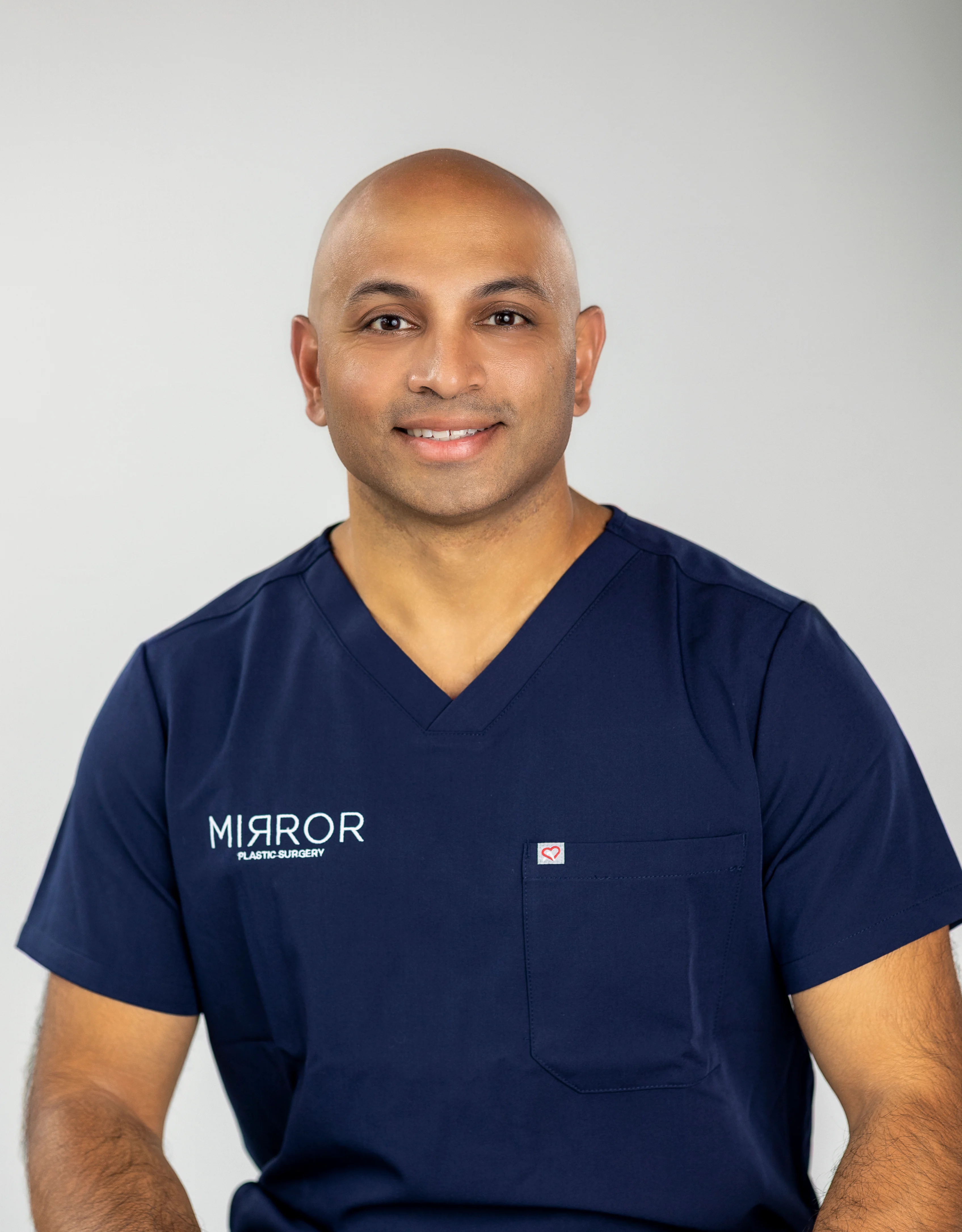
As an advisory board member for Merz Aesthetics and Tiger Aesthetics, Dr. Chandawarkar stays closely connected to emerging data on injectable safety and innovation. His supplier-neutral philosophy keeps product recommendations grounded in patient anatomy and clinical evidence, without commercial pressure.
Mirror Plastic Surgery follows a concierge model, with consultations lasting up to an hour. These visits include a detailed assessment of facial anatomy, medical history, immune status, and prior treatments. The practice limits daily procedures so each patient receives focused, personalized care before, during, and after injections. This structure directly supports safer outcomes through thorough screening, precise technique, and attentive follow-up.
Patients considering fillers for the first time or seeking help after previous treatments can expect an evidence-based, safety-focused approach. Schedule a personalized injectable consultation at Mirror Plastic Surgery in St. Petersburg, FL, or call 727-361-6515 to speak with the team.
Conclusion
Research consistently shows that granulomas and nodules after facial dermal fillers are uncommon. Delayed complications can appear months to years after injection and may involve biofilms, immune triggers, or filler properties. Most nodules, especially those related to HA fillers, resolve over time, and the majority of all adverse events remain mild and self-limited. When treatment becomes necessary, options such as hyaluronidase, intralesional corticosteroids, antibiotics, and, rarely, surgery provide effective management.
Choosing a qualified, board-certified provider remains the most powerful step patients can take to support safe outcomes. A surgeon with advanced anatomical expertise can inject thoughtfully, diagnose accurately, and manage complications when they occur. If you have noticed a lump after filler treatment or want to review your injectable options with a safety-focused surgeon, Mirror Plastic Surgery offers comprehensive, evidence-based care.
Disclaimer: Results may vary from person to person. Editorial content, before and after images, and patient testimonials do not constitute a guarantee of specific results.
1 Results may vary from person to person. Editorial content, before and after images, and patient testimonials do not constitute a guarantee of specific results.