Likelihood of Asymmetry and Risks After Lower Blepharoplasty
Content
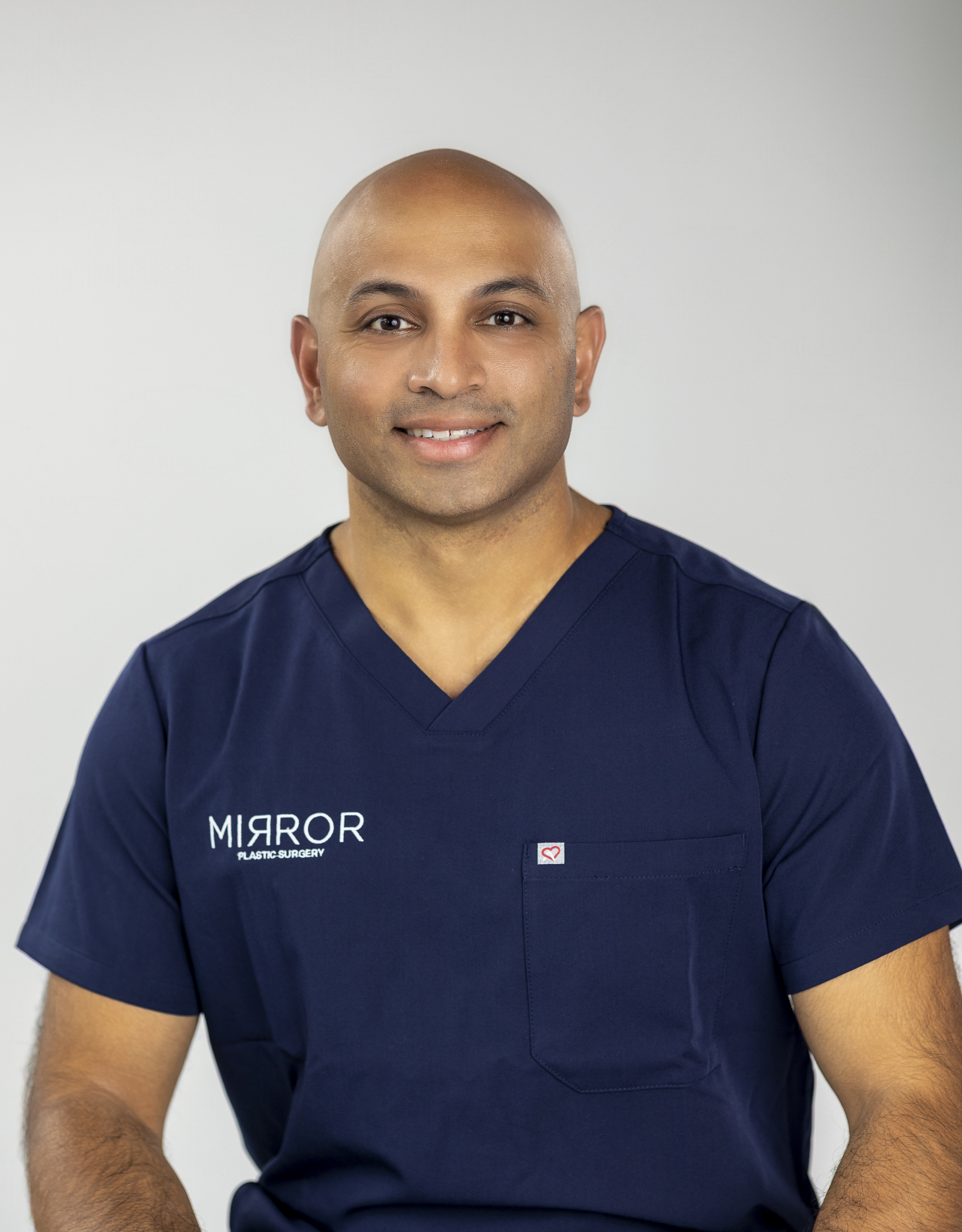
Written by: Dr. Akash Chandawarkar, Board Certified Plastic Surgeon, Mirror Plastic Surgery | Last updated: June 27, 2026
Key Takeaways
Lower blepharoplasty treats under-eye bags and restores smoother contours through either a transcutaneous or transconjunctival approach, each with a distinct risk profile.
Most complications such as asymmetry, dry eye, and chemosis are temporary and resolve within weeks to months, while serious permanent issues remain uncommon with experienced surgeons.1
The transconjunctival approach generally carries lower risks of ectropion and eyelid retraction than the transcutaneous method, especially when skin removal is not required.1
Patients should plan for at least two weeks of social downtime, understand healing timelines up to 12 months, and recognize that revision surgery is usually deferred until swelling fully resolves.1
Selecting an experienced, board-certified surgeon is the most important factor in minimizing risks; schedule your personalized consultation at Mirror Plastic Surgery to discuss your anatomy and safety priorities.
Modern Lower Blepharoplasty Standards and Safety Priorities
Lower blepharoplasty ranks among the most frequently performed facial rejuvenation procedures in the United States. Technique evolution now favors volume preservation and fat repositioning instead of aggressive fat removal, because over-resection strongly correlates with complications and revision surgery. Contemporary safety standards call for detailed preoperative ocular assessment, conservative tissue handling, and surgeon-level knowledge of eyelid anatomy that extends beyond surface aesthetics. Patient selection, overall health, and adherence to pre- and postoperative instructions play equally important roles in final outcomes.
Asymmetry refers to a visible difference in the position, contour, or volume of the two lower eyelids after surgery. This difference may be temporary and driven by uneven postoperative swelling, or persistent and related to healing patterns, fat removal, or preexisting anatomical variation.
Ectropion is an outward turning of the lower eyelid margin away from the globe. It can result from excessive skin removal, scar contracture, or inadequate support of the lid-cheek complex, and ranges from mild rounding of the outer canthus to full eversion that requires surgical correction.
Eyelid retraction describes downward displacement of the lower lid margin that exposes sclera below the iris. It shares causes with ectropion and is a recognized risk of the transcutaneous approach when the anterior lamella is shortened.
Chemosis is edema of the bulbar conjunctiva, which appears as a clear, gelatinous swelling on the eye surface. It occurs more often when lower blepharoplasty is combined with upper eyelid surgery or a facelift and typically resolves over a few weeks, although cases lasting beyond two to three weeks benefit from targeted topical treatment.
Retrobulbar hemorrhage is a rare but vision-threatening accumulation of blood behind the globe that can elevate intraorbital pressure and compress the optic nerve. It represents a surgical emergency that requires immediate intervention and is among the most serious risks associated with any periorbital procedure.
Quantified Complication Data: Temporary Versus Permanent Risks
Once these key complications are defined, their frequency and usual timelines for resolution provide helpful context for decision-making. The table below summarizes documented complication rates and expected resolution windows drawn from published clinical data. Evidence strength varies, because prospective controlled trials specific to lower blepharoplasty remain limited, and figures reflect pooled or observational data.
Resolves within several months, permanent in rare cases
Note: Incidence figures derive from observational and pooled data. Individual risk varies with technique, surgeon experience, patient anatomy, and health status. Retrobulbar hemorrhage is not included in the table because its incidence is too rare and variably reported across sources to cite a reliable pooled figure; it is addressed in the technique comparison below.
Comparing Transcutaneous and Transconjunctival Lower Eyelid Approaches
The transcutaneous approach places an incision just below the lash line and provides direct access to skin, muscle, and fat. This access comes with a higher risk of anterior lamellar shortening, which is the primary mechanism behind ectropion and eyelid retraction. Scar contracture along the external incision can pull the lid margin downward over time, particularly in patients with poor lower lid tone or preexisting negative vector anatomy.
The transconjunctival approach places the incision inside the eyelid, which eliminates an external scar and avoids disruption of the anterior lamella. This technique is generally associated with lower rates of ectropion and retraction because the structural support of the lower lid remains intact. It works best for patients whose main concern is fat herniation without significant excess skin. When skin removal is also needed, a separate pinch excision or laser resurfacing can be added to the transconjunctival approach, preserving its safety advantages while addressing surface laxity.
Excessive skin removal during blepharoplasty can cause inability to fully close the eyelids, but this complication is rare when a skilled surgeon uses a cautious approach to tissue excision. Retrobulbar hemorrhage risk exists with both approaches whenever fat is manipulated and is reduced through meticulous hemostasis and avoidance of aggressive fat excision. Surgeon volume and anatomical expertise remain the most consistent predictors of technique-specific outcomes across both approaches.
Healing Timelines After Lower Blepharoplasty
1 Week: Swelling and bruising peak at 48–72 hours postoperatively before gradually improving, and most visible bruising and swelling improve substantially within 10–14 days.1 Asymmetry at this stage almost always reflects uneven swelling rather than a structural problem.
1 Month: At 3–4 weeks, most patients appear natural to casual observers, though minor swelling or asymmetry may still fluctuate, especially in the mornings.1 The early asymmetry described in the complication table often remains in this window but usually continues to improve. Dry-eye symptoms, if present, are typically managed with lubricating drops at this stage.
6–12 Months: Final results are considered established during this period. Asymmetry that persists after swelling resolution may require minor revision surgery and is typically addressed in the revision timeframe described below.
Revision Rates and Primary Reasons for Secondary Surgery
Common reasons patients seek revision blepharoplasty include overcorrection (excessive removal leading to hollowing or lagophthalmos), undercorrection (persistent puffiness or excess skin), asymmetry, hypertrophic scarring, and functional problems such as ectropion, entropion, or dry eyes.
Revision blepharoplasty carries increased risks and diminishing returns compared with primary procedures because of scar tissue formation and disrupted blood supply from prior operations. Functional complications including inability to fully close the eyes or severe dry-eye symptoms may justify earlier revision before the standard 6–12 month waiting period.
Practical Planning Tips for Lower Blepharoplasty Patients
Patients researching lower blepharoplasty benefit from entering consultations ready to discuss baseline tear production, lower lid tone (snap test and distraction test results), and any history of dry eye or contact lens use. These factors directly influence technique selection and postoperative risk. Helpful questions include the surgeon’s preferred approach for the patient’s specific anatomy, the number of lower blepharoplasty procedures performed annually, and the practice’s protocol for managing complications such as chemosis or early ectropion signs.
Recovery planning should include a minimum of two weeks of social downtime and avoidance of strenuous activity. Patients should understand that rapidly increasing swelling on one side, sudden vision changes, or bleeding that does not slow with gentle pressure require prompt contact with the surgical team. Strict adherence to a surgeon’s pre- and post-operative restrictions on medications, supplements, cigarettes, and alcohol reduces the risk of serious complications such as poor healing or infection.
As discussed earlier, surgeon selection remains the single most modifiable risk factor, with board certification and consistent outcomes serving as primary evaluation criteria. Most complications can be avoided by choosing a board-certified facial plastic surgeon with excellent safety records and a conservative approach.
Expert Perspective: Dr. Akash, MD Harvard-MIT HST Medical Degree | Johns Hopkins Integrated Plastic Surgery Residency | MEETH Aesthetic Surgery Fellowship | Named to Newsweek’s America’s Best Plastic Surgeons 2025 for two consecutive years
Dr. Akash performs lower blepharoplasty with a safety-first, anatomy-driven philosophy and often dedicates up to an hour per consultation to assess lower lid tone, tear-film health, and orbital anatomy before recommending a technique. Mirror Plastic Surgery limits surgical volume to one to two procedures per day, which preserves undivided perioperative focus on each patient.
Published complication rates for lower blepharoplasty vary considerably across studies because of differences in patient populations, technique definitions, follow-up duration, and how complications are classified and reported. Prospective randomized data comparing transcutaneous and transconjunctival approaches with standardized outcome measures remain limited. Surgeon volume effects are widely acknowledged in the clinical community but remain difficult to quantify precisely in published literature. Individual healing variability, influenced by genetics, skin quality, age, and systemic health, means that population-level statistics serve as directional guides rather than individual predictions. Patients with preexisting dry eye, negative vector orbits, or prior periorbital surgery carry baseline risks that may exceed published averages.
How Modern Techniques and Concierge Care Improve Safety
The shift toward fat repositioning instead of excision, the integration of midface support techniques, and the growing use of preoperative imaging and ocular surface assessment reflect a maturing understanding of lower eyelid anatomy. Practices that prioritize anatomical knowledge, conservative tissue handling, and thorough preoperative evaluation tend to achieve lower revision rates and higher patient satisfaction. The concierge model, in which surgical volume is deliberately limited to preserve perioperative attention, directly addresses conditions under which many preventable complications arise, including rushed assessment, inadequate patient selection, and insufficient postoperative monitoring.
Visible asymmetry at one month most often stems from uneven postoperative swelling rather than a structural surgical problem. Swelling after lower blepharoplasty rarely resolves symmetrically, and one side frequently lags behind the other by days or weeks. Mild asymmetry in the first three months is expected and typically resolves without intervention as edema continues to dissipate. Patients who notice one side appears more swollen, feels firmer, or shows more bruising should document the change with photographs and report any rapidly worsening asymmetry, pain, or vision changes to their surgeon immediately, because these signs can indicate a complication that needs prompt evaluation. Asymmetry that persists and remains stable after swelling has resolved is more likely to reflect a structural difference and may be a candidate for minor revision after full healing is confirmed.
Long-Term Side Effects of Lower Blepharoplasty
Most side effects after lower blepharoplasty are temporary. Long-term or permanent side effects remain uncommon when an experienced, board-certified surgeon uses conservative tissue-handling principles. Potential long-term issues include persistent dry eye in patients with preexisting tear-film dysfunction, ectropion or eyelid retraction from excessive skin removal or scar contracture, permanent changes in lower lid position, and, very rarely, permanent numbness or vision changes. Patients with negative vector orbital anatomy, poor lower lid tone, or a history of prior periorbital surgery carry a higher baseline risk for long-term complications and should discuss these factors explicitly during consultation.
Risk Differences Between Transcutaneous and Transconjunctival Approaches
The transcutaneous approach, which uses an external incision below the lash line, provides access to skin, muscle, and fat but carries a higher risk of ectropion and eyelid retraction because it can shorten the anterior lamella of the eyelid. The transconjunctival approach, placed inside the eyelid, avoids an external scar and preserves the structural support of the lower lid, which generally results in lower rates of lid malposition. The transconjunctival approach works best for patients whose primary concern is fat herniation without significant excess skin. When skin laxity is also present, a conservative external pinch excision or laser resurfacing can be added. The optimal technique depends on individual anatomy, skin quality, lid tone, and the surgeon’s assessment, rather than a universal preference for one approach.
Timing and Limits of Revision Surgery
Most surgeons recommend waiting a minimum of six to twelve months after primary lower blepharoplasty before pursuing revision surgery, which allows swelling to fully resolve, scar tissue to mature, and final results to stabilize. Exceptions include functional emergencies such as inability to close the eye or severe corneal exposure, which may require earlier intervention. Common indications for revision include persistent asymmetry, ectropion, hollowing from over-resection, or residual fat herniation from under-resection. There is no fixed limit on the number of revisions, but each successive procedure becomes more technically demanding because of scar tissue and altered tissue planes, and the risk-benefit ratio worsens with each additional operation. Patients considering revision should seek a surgeon with specific experience in secondary eyelid surgery.
Steps to Reduce Complication Risk
Risk reduction starts with thorough preoperative evaluation that includes assessment of tear production, lower lid tone, and orbital anatomy. Patients should disclose all medications, supplements, and health conditions, because many common substances, including aspirin, NSAIDs, vitamin E, fish oil, and herbal supplements, increase bleeding risk. Smoking impairs wound healing and should be stopped well in advance of surgery. After surgery, strict adherence to the surgeon’s instructions regarding activity restriction, head elevation, cold compresses, and prescribed eye drops significantly reduces the likelihood of complications. Attending all follow-up appointments allows early identification and management of issues such as chemosis, early ectropion signs, or dry-eye progression before they become entrenched problems.
Summary and Next Steps
The overall complication rate for lower blepharoplasty is generally low, and the large majority of complications are mild, temporary, and self-resolving.1 Asymmetry in the first three months most often reflects swelling rather than a structural problem. Serious permanent complications such as ectropion, retraction, lagophthalmos, and retrobulbar hemorrhage remain uncommon and are most effectively prevented through conservative technique, thorough preoperative assessment, and careful surgeon selection. Technique choice between transcutaneous and transconjunctival approaches should be tailored to individual anatomy. When needed, revision surgery is usually deferred until six to twelve months postoperatively, except in functional emergencies.
Patients who approach consultation with specific questions about technique rationale, complication management protocols, and surgeon volume are better positioned to decide whether a practice’s philosophy aligns with their safety priorities. Mirror Plastic Surgery’s concierge model, which limits surgery to one to two cases per day and allows up to an hour per consultation within a safety-first clinical hierarchy, is designed for patients who value that level of rigor.
Request your comprehensive anatomical assessment at Mirror Plastic Surgery in St. Petersburg, Florida. Dr. Akash, who completed Harvard-MIT training, a Johns Hopkins residency, and a MEETH fellowship and was named to Newsweek’s America’s Best Plastic Surgeons 2025 for two consecutive years, will provide transparent, evidence-based guidance on your lower blepharoplasty options.
Medical Disclaimer: The information provided in this article is intended for general educational purposes only and does not constitute medical advice, diagnosis, or treatment. Individual risks, outcomes, and timelines vary based on patient anatomy, health status, surgical technique, and surgeon experience. Consult a board-certified plastic surgeon for a personalized evaluation before making any surgical decisions.
1 Results may vary from person to person. Editorial content, before and after images, and patient testimonials do not constitute a guarantee of specific results.