

Written by: Ellie Pranckevicius, FNP-BC, Aesthetic Nurse Practitioner & Aesthetic Injector | Facial Restoration & Regenerative Injectable Specialist, Mirror Plastic Surgery | Last updated: August 6, 2026
Key Takeaways
- Natural facial volume restoration works best when treatment rebuilds deep structural support at the bone level instead of adding superficial bulk.1
- A comprehensive top-to-bottom facial assessment that includes emotional goals and anatomy sets the stage for balanced, natural results.
- Deep periosteal bolus injections at key bony landmarks combined with cannula vector lifts create foundational support and help lower vascular risk.1
- Product choice between hyaluronic acid fillers and biostimulatory agents should reflect anatomy, timing, and reversibility needs rather than brand loyalty.
- Patients who want natural facial volume restoration can schedule a personalized consultation with Ellie Pranckevicius at Mirror Plastic Surgery for an anatomy-first treatment plan.
Ellie’s Full-Face Assessment Protocol for Natural Volume Restoration
Every natural-looking filler result starts with a structured pre-treatment assessment. At Mirror Plastic Surgery, Ellie Pranckevicius performs a comprehensive top-to-bottom evaluation that can last up to an hour and covers both emotional goals and anatomical realities. This mirrors the framework outlined in a 2026 Dermatologic Surgery review, which states that whole-face assessment must account for regional facial aging patterns that vary across sex, age, and ethnicity to support natural outcomes and avoid overcorrection.
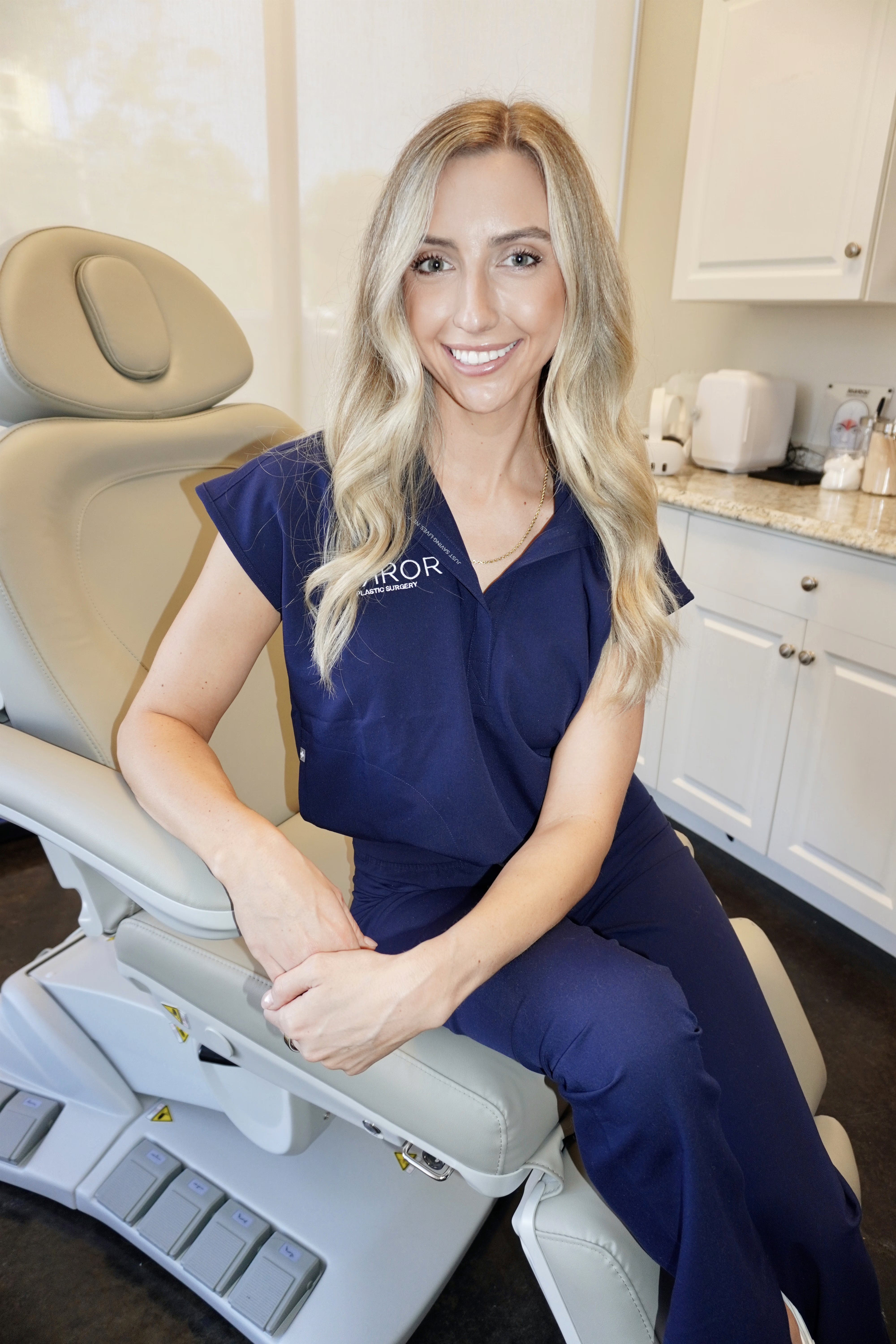
The protocol uses symptom-based severity scales, such as grading temporal hollowing, and structured diagnostic tools comparable to the Galderma Facial Assessment Scale (FASTM), which improves clinician-patient communication and individualized treatment planning. Ellie focuses first on restoring inherent facial characteristics before adding volume. She treats the upper, middle, and lower face as a continuous system rather than isolated zones. Once this assessment identifies which structures need support, treatment planning moves to the deepest layer where skeletal support begins.
Deep Periosteal Bolus Landmarks for Foundational Support
Supraperiosteal injections are preferred for structural lift and projection in areas such as the piriform fossa, zygomatic arch, and mandibular angle because they restore foundational support with lower vascular risk when performed on bone with aspiration. The 2026 L-V (lifting and V-shaped contouring) approach by Wu et al. turns this concept into a clear, sequenced protocol.
- Begin with deep supraperiosteal bolus injections at bony landmarks such as the temple, zygomatic arch, nasal base, and chin, using high G′ filler for foundational skeletal support.
- Reinforce anchoring structures with targeted ligament and fascia injections, using high G′ fillers at the zygomatic, orbital, and mandibular retaining ligaments to reposition sagging tissues.
- Refine the surface with superficial placement of lower G′ fillers to smooth dynamic grooves while preserving natural expression.
Safety at the jawline requires specific checkpoint discipline because the facial artery’s path creates distinct high-risk and lower-risk zones. The facial artery crosses the mandible periosteally at the antegonial notch, which makes this crossing a critical safety checkpoint for deep periosteal jawline bolus injections. In contrast, the posterior jawline near the gonial angle is a relatively avascular zone suitable for periosteal bolus injection. Even in safer zones, mandatory aspiration and slow, low-pressure delivery remain essential at every deep landmark.
Cannula Vector Lifts for Midface and Jawline Contouring
After foundational periosteal support is in place, Ellie uses blunt-tip cannulas to distribute volume along precise lifting vectors in the midface and jawline. Evidence from split-face randomized controlled trials and cohort studies shows that cannulas can reduce bruising compared with needles while achieving similar aesthetic outcomes.
Cannula vector strategy follows the anatomical descent pattern of aging, which tends to move lateral to medial and superior to inferior. Studies suggest that needle injections can cause filler to shift between tissue planes, while cannula injections provide more stable placement within a single plane. This stability is especially valuable in the midface subcutaneous layer, where unintended redistribution can widen facial contours over time. Beyond preventing aesthetic migration, cannulas also help reduce vascular risk in high-risk zones because their blunt tip is less likely to penetrate vessel walls than a sharp needle, especially when paired with low plunger pressure and small aliquots.
Micro-Droplet Blending for Smooth Zone Transitions
Micro-droplet technique connects deep structural boluses with the skin surface and creates smooth transitions between facial zones. Product rheology must match the target plane, and natural-looking volume restoration requires matching product rheology and injection plane to the targeted anatomical layer rather than using a uniform technique across the face.
Plane accuracy matters because typical 30° to 45° needle insertion angles place filler almost always into the subcutaneous tissue, while insertion angles of 7° to 12° are required to consistently deposit filler within the dermis. For superficial dynamic areas such as nasolabial and marionette lines, lower G′ fillers help preserve natural expression while softening surface irregularities without adding bulk.
Temple and Chin: High-Impact Zones with Higher Risk
Temples and chin often receive less attention, yet they strongly influence overall facial balance. Both zones carry higher vascular risk and require precise anatomical understanding.
In the temple, a 2026 morphometric CT angiography study by Soyal et al. identified high-risk zones for the superficial temporal artery and zygomatico-orbital artery and recommended injections in the periosteal plane. The same study reported that aging causes lateral and superficial displacement of the superficial temporal artery and middle temporal vein, which heightens vascular risk during blind periosteal injections in older patients. This shift affects exactly the group most likely to seek temple volume restoration.
At the chin, the submental artery, which branches from the facial artery and runs along the anterior mandible, is the vessel most often involved in chin-area vascular occlusions during deep injections. Ultrasound guidance or color Doppler mapping before injection in these high-risk corridors now represents the standard of care for complex cases.
Choosing Between HA Fillers and Biostimulatory Products
Product selection functions as a clinical decision, not a brand preference. Ellie bases this choice on the pattern of volume loss, skin quality, timing for visible results, and the need for reversibility.
Biostimulatory agents including Sculptra (PLLA) and Radiesse (CaHA) act as scaffolds that trigger fibroblast recruitment and neocollagenesis, so long-term visible volume derives from the patient’s own deposited collagen. Sculptra produces sustained facial volume correction up to 2+ years via the patient’s own collagen, while Radiesse provides immediate volume plus collagen stimulation lasting 12–18 months.1
Hybrid protocols increasingly used in 2026 combine immediate HA structure with diffuse biostimulator collagen building. This approach lets Ellie address both acute volume needs and long-term tissue quality within one plan. Her supplier-neutral position at Mirror Plastic Surgery keeps product selection focused on anatomy and goals rather than brand quotas.
Safety Checkpoints and Long-Term Filler Maintenance
Vascular occlusion represents the most serious non-surgical filler complication, even though it remains rare. Ellie follows a strict set of prevention checkpoints during every treatment.
- Pre-injection topographic assessment and, in high-risk zones, color Doppler or ultrasound mapping
- Mandatory aspiration at deep periosteal sites near named vessels
- Slow, low-pressure delivery in aliquots of ≤0.1 mL per pass
- Cannula preference in superficial high-risk planes such as the nasolabial fold, tear trough, and temple subcutaneous layer
- Continuous visual and tactile monitoring for blanching, mottling, or patient-reported pain during injection
When vascular compromise is suspected, first-line management follows the DeLorenzi high-dose pulsed hyaluronidase protocol of 500–1500 IU delivered into and around the ischemic territory every 60 minutes until reperfusion, supported by warm compresses, a 300 mg aspirin loading dose, and immediate ophthalmology referral for any visual symptoms.
Post-treatment maintenance follows a “less-is-more” philosophy. Final dermal filler results become visible at 2–4 weeks, and patients should schedule a follow-up review appointment at the two-to-four-week interval to assess settling and perform any necessary adjustments.1 Strenuous exercise should typically be avoided for 24–48 hours after dermal filler treatment to minimize swelling, bruising, and risk of filler movement. Long-term, daily broad-spectrum SPF 30 or higher is recommended to help prevent UV-induced filler degradation.
Zone-Goal Reference Table
| Treatment Zone | Primary Goal | Preferred Depth | Product Category |
|---|---|---|---|
| Temple | Restore lateral facial frame and reduce skeletonization | Supraperiosteal; ≥2 cm from lateral orbital rim | High G′ HA or biostimulatory (Sculptra/Radiesse) |
| Zygomatic arch / Malar | Midface projection and fat-pad support | Supraperiosteal (Layer 5) | Intermediate G′ HA (G′ ~579 Pa) for apple cheek |
| Infraorbital / Tear trough | Hollow correction and reduced shadowing | Deep SOOF beneath orbicularis; ultrasound-confirmed | Low-hydrophilic HA; cannula preferred |
| Nasolabial / Marionette | Soften grooves while preserving expression | Subcutaneous (Layer 2); cannula in superficial plane | Low G′ HA (G′ ~117 Pa) |
| Jawline / Gonial angle | Definition and jowl reduction | Periosteal at gonial angle; cannula at mid-mandibular body | High G′ HA or Radiesse |
| Chin | Projection and lower-face balance | Deep periosteal; caution for submental artery | High G′ HA; needle for precision bolus |
Conclusion: Putting the Techniques Together for Natural Results
The most effective dermal filler techniques for natural facial volume restoration share a common foundation of anatomy-first assessment, depth-appropriate product placement, zone-specific vector strategies, and a disciplined complication-prevention protocol. No single technique or product achieves natural results alone. As discussed in the assessment framework, this diversity-informed approach merges clinical precision with individualized beauty goals.
Patients considering full-face restorative filler treatment benefit from a provider who performs a thorough pre-treatment assessment, selects products based on individual anatomy rather than brand affiliation, and maintains a written vascular emergency protocol on-site. Ellie Pranckevicius and the team at Mirror Plastic Surgery apply these standards to every patient in the St. Petersburg and Tampa Bay area.
Disclaimer: Results may vary from person to person. Editorial content, before and after images, and patient testimonials do not constitute a guarantee of specific results.
Start Your Anatomy-First Consultation with Ellie
Ellie Pranckevicius is currently welcoming new clients seeking full-face restorative filler treatments at Mirror Plastic Surgery in St. Petersburg, Florida. Every new patient receives a comprehensive top-to-bottom assessment, not a rushed appointment, before any treatment is recommended.
Frequently Asked Questions
What makes deep periosteal filler placement safer than superficial injection for volume restoration?
Deep periosteal placement positions filler directly on bone, beneath the layers where major facial arteries branch and travel toward the midline. Because the primary vascular structures run in the superficial subcutaneous and subdermal planes, injecting at the periosteal level with proper aspiration and slow delivery significantly reduces the risk of inadvertent intravascular placement. Filler at this depth also provides true structural support by rebuilding the bony scaffolding that aging erodes rather than simply adding bulk to soft tissue. This approach produces longer-lasting, more anatomically coherent results and avoids the puffiness associated with superficial volume addition.1 At Mirror Plastic Surgery, Ellie Pranckevicius combines periosteal bolus technique with real-time anatomical assessment and, when indicated, ultrasound or Doppler mapping to confirm safe placement before injection.
How does Ellie decide between hyaluronic acid fillers and biostimulatory agents like Sculptra or Radiesse?
Ellie bases this decision on several patient-specific factors, including the size and distribution of volume loss, skin quality, the patient’s timeline for visible results, and whether reversibility is a priority. Hyaluronic acid fillers deliver immediate, precise volume and can be dissolved with hyaluronidase if needed, which makes them well-suited for localized corrections such as the tear trough, lips, or acute asymmetry where exact placement and adjustability matter. Biostimulatory agents like Sculptra and Radiesse work better for diffuse volume loss across larger areas such as the temples, cheeks, and jawline because they stimulate the patient’s own collagen production over months and create gradual, tissue-integrated results that can last two years or more. In many full-face restorative plans, Ellie uses a hybrid approach with biostimulatory agents for broad foundational rebuilding and hyaluronic acid fillers for precise contouring in specific zones. Because Mirror Plastic Surgery is supplier-neutral, product selection stays focused on what best fits each patient’s anatomy.
What should I expect during recovery after a full-face filler treatment?
Most patients experience mild redness, swelling, tenderness, and occasional bruising in the days after treatment. Swelling usually peaks at 24–48 hours and resolves within one to two weeks, although some areas continue to soften and settle during that time. Final results are generally easier to assess at about two weeks, so Mirror Plastic Surgery schedules a follow-up review at that point to evaluate settling and make any refinements. During the first 48 hours, patients should avoid vigorous exercise, alcohol, heat exposure, retinoids, and NSAIDs unless medically necessary. Full exercise, including resistance training and cardio, should wait about one week. Sleeping with the head elevated for the first few nights and avoiding face-down positions helps prevent product displacement while it integrates with surrounding tissue. Daily broad-spectrum SPF 30 or higher is recommended long-term because UV exposure accelerates filler breakdown. Patients should contact their provider immediately if they notice skin blanching, bluish or mottled discoloration, worsening pain, or any vision changes, since these signs can indicate a vascular complication that needs urgent treatment.
Why does Mirror Plastic Surgery emphasize a full-face assessment before any filler treatment?
Isolated, single-area filler injections without a whole-face evaluation often create imbalanced or unnatural results. For example, adding cheek volume without addressing temple hollowing can make the face appear disproportionate. A full-face assessment lets the injector understand how each zone relates to the others, identify the root anatomical causes of aging rather than just surface symptoms, and build a sequenced plan that addresses structure before volume. Ellie’s top-to-bottom protocol also explores the emotional drivers behind a patient’s goals, which shapes treatment priorities and supports realistic expectations. This approach reflects Mirror Plastic Surgery’s core philosophy of safety first, function second, and aesthetics third. The result is a treatment plan tailored to each patient’s anatomy, aging pattern, and long-term goals rather than a standardized menu of procedures.
Is there a risk of looking overfilled, and how does Ellie prevent it?
The overfilled appearance usually results from superficial placement that adds bulk without structural support, repeated volume addition to a single zone without pan-facial balance, use of high-hydrophilic products in areas prone to water retention, or lack of planning for how filler redistributes within tissue compartments over time. Ellie prevents this outcome through several safeguards. The full-face assessment identifies which areas truly need volume and which need lifting or skin quality improvement, and sometimes the honest answer is that a patient does not yet need a specific treatment. Product selection matches rheology to anatomical layer and avoids high-water-attracting fillers in zones where swelling would be obvious. The sequenced injection protocol builds deep structural support before superficial refinement so that volume is added only where the anatomy calls for it. Mirror Plastic Surgery’s “less-is-more” philosophy favors minor, planned corrections over aggressive single-session volume loading, with results reassessed at two weeks before any additional product is considered.
1 Results may vary from person to person. Editorial content, before and after images, and patient testimonials do not constitute a guarantee of specific results.